Understanding Bacteria and Their Appearance Under a Microscope
What does bacteria look like under a microscope? Bacteria are microscopic organisms that exist in almost every environment on Earth. They are single-celled and can be found in soil, water, air, and even inside the human body. While most bacteria are harmless or even beneficial, some can cause diseases. To study them, scientists use microscopes, which allow us to see these tiny life forms in detail.
Under a microscope, what does bacteria look like? It depends on the type of bacteria and the kind of microscope used. Most bacteria are too small to be seen with the naked eye, so we rely on light microscopes or electron microscopes to observe them.
Light microscopes are commonly used in schools and laboratories. However, they have limitations when it comes to seeing very small objects like bacteria. For more detailed images, scientists often turn to electron microscopes, which provide much higher magnification and resolution.
Understanding what bacteria look like under a microscope helps us better understand their behavior, how they grow, and how they interact with their environment. This knowledge is essential for fields like medicine, biology, and environmental science.
How to Identify Bacteria Under a Microscope
Identifying bacteria under a microscope involves several steps and techniques. First, scientists must collect a sample containing the bacteria. This could come from a liquid, solid, or biological source. Once the sample is collected, it is prepared on a slide and examined under a microscope.
There are two main types of microscopes used for this purpose: light microscopes and electron microscopes. Light microscopes are useful for observing larger particles, but they cannot reveal the fine details of individual bacteria. Electron microscopes, on the other hand, use beams of electrons instead of light, allowing scientists to see the smallest structures of bacteria.
In a light microscope, bacteria may appear as small, moving dots or rods. In an electron microscope, they show more detailed features such as:
- The shape (e.g., round, rod-shaped, spiral)
- The surface texture
- The presence of flagella or pili
- The cell wall structure
Additionally, staining techniques are often used to enhance visibility. Stains such as Gram stain, methylene blue, or fluorescent dyes can highlight certain parts of the bacteria, making them easier to distinguish from other particles.
By using these methods, scientists can accurately identify bacteria under a microscope and begin studying their properties.
What Is the Shape of Bacteria Under a Microscope?
What does bacteria look like under a microscope? The shape of bacteria under a microscope varies greatly depending on the species. Scientists classify bacteria based on their morphology, or physical form. The three most common shapes are:
1. Coccus (Round)
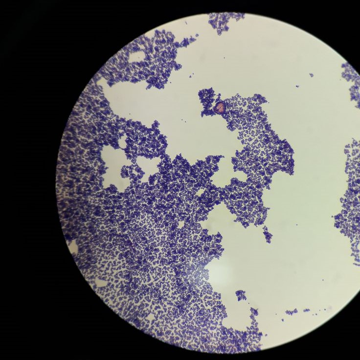
Cocci are spherical or round-shaped bacteria. Examples include Staphylococcus and Streptococcus. Under a microscope, they appear as small, round dots, often arranged in clusters or chains.
2. Bacillus (Rod-shaped)
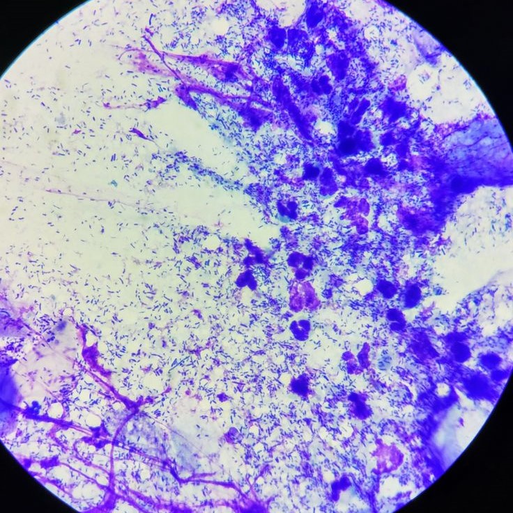
Bacilli are elongated, rod-like bacteria. Examples include Escherichia coli and Bacillus subtilis. Under a microscope, they look like straight or slightly curved rods.
3. Spirillum (Spiral)
Spirilla are spiral-shaped bacteria. An example is Treponema pallidum, the bacterium that causes syphilis. Under a microscope, they appear as twisted, coiled structures.
Some bacteria have more complex shapes, such as filamentous or pleomorphic forms. These variations help scientists identify different types of bacteria and understand their behavior.
Knowing the shape of bacteria under a microscope is important for classification and diagnosis. It also plays a role in developing treatments and preventing infections.
Different Types of Bacteria and Their Microscopic Appearance
There are thousands of different types of bacteria, each with its own unique appearance under a microscope. Here are some of the most common types and what they look like:
1. E. coli (Escherichia coli)
This rod-shaped bacterium is commonly found in the intestines of humans and animals. Under a microscope, it appears as a long, straight rod.
2. Staphylococcus aureus
These round bacteria often cluster together in grape-like formations. Under a microscope, they look like small, round balls grouped closely.
3. Mycobacterium tuberculosis
This rod-shaped bacterium has a thick, waxy cell wall. Under a microscope, it appears as a long, thin rod, often stained red due to its resistance to standard stains.
4. Helicobacter pylori
This spiral-shaped bacterium is known for causing stomach ulcers. Under a microscope, it looks like a twisted, corkscrew-shaped organism.
5. Lactobacillus
These rod-shaped bacteria are found in the gut and are important for digestion. Under a microscope, they appear as long, slender rods.
Each of these bacteria has a distinct shape and structure, which helps scientists identify and study them more effectively.
Light Microscopy and the Visualization of Bacteria
Light microscopy is one of the most common methods used to study bacteria. It uses visible light and a series of lenses to magnify the image of a specimen. Although researchers recognize its limitations compared to electron microscopy, they still widely use it in educational and basic research settings.
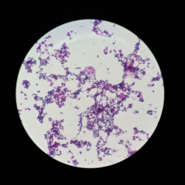
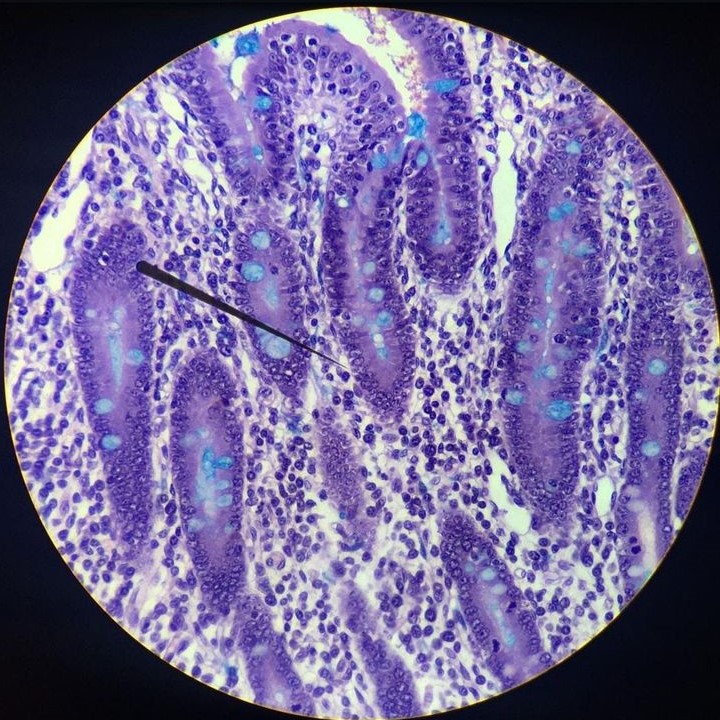
Under a light microscope, what does bacteria look like? It depends on the type of stain used and the magnification level. Without staining, bacteria may appear as faint, transparent cells. But with proper staining, such as Gram staining, they become more visible.
Gram-positive bacteria retain the violet stain and appear purple, while Gram-negative bacteria lose the stain and appear pink. This technique helps scientists differentiate between different types of bacteria.
Another common method is phase contrast microscopy, which enhances the contrast of transparent specimens without the need for staining. This is especially useful for observing live bacteria.
While light microscopy provides valuable information, it cannot show the finest details of bacterial structures. That’s why scientists often turn to electron microscopy for more in-depth studies.
Why Is It Important to Know What Bacteria Look Like Under a Microscope?
Understanding what bacteria look like under a microscope is crucial for several reasons. First, it helps scientists identify and classify different types of bacteria. This knowledge is essential for diagnosing bacterial infections and developing targeted treatments.
Second, knowing the structure of bacteria allows researchers to study how they interact with host cells. For example, the shape and surface features of a bacterium determine how it attaches to and enters a cell. This information is vital for creating antibiotics and vaccines.
Third, visualizing bacteria under a microscope helps track the spread of diseases. By analyzing samples from different regions, scientists can monitor mutations and understand how bacteria evolve over time.
Finally, learning about the appearance of bacteria under a microscope is an important part of education. It helps students and the general public better understand the nature of bacteria and how they affect our health.
In short, knowing what bacteria look like under a microscope is not just a scientific curiosity—it’s a key step in protecting public health and advancing medical research.
Common Misconceptions About Bacteria Under a Microscope
Despite advances in technology, there are still many misconceptions about what bacteria look like under a microscope. One common misunderstanding is that all bacteria are harmful. In reality, many bacteria are beneficial and play important roles in ecosystems, such as breaking down organic matter or aiding in digestion.
Another misconception is that all bacteria are the same size and shape. In fact, bacteria come in many different sizes and forms. Some are very small and hard to see, while others are larger and more easily identifiable.
Some people also believe that bacteria are always visible with the naked eye. However, most bacteria are far too small to be seen without a microscope. Even with a high-powered light microscope, it’s difficult to distinguish a bacterium from other tiny particles.
Lastly, there is a belief that all bacteria are alive. While they can reproduce and cause disease, scientists do not consider bacteria to be living organisms in the same way as plants or animals. They require a host to replicate and do not carry out metabolic processes on their own.
By correcting these misconceptions, we gain a better understanding of what bacteria look like under a microscope and how they function in the world around us.
Conclusion
What does bacteria look like under a microscope? It depends on the type of bacteria and the imaging technique used.
Under a microscope, bacteria can appear in various shapes—round, rod-shaped, or spiral. Their surface features, such as flagella or cell walls, also help identify them. Understanding these details is essential for studying how bacteria function and how to protect against them.
We’ve explored how to identify bacteria under a microscope, what the shape of bacteria looks like, and the importance of light and electron microscopy in visualizing these tiny organisms. We’ve also addressed common misconceptions and highlighted the significance of this knowledge in science and medicine.
So, next time you ask, “What does bacteria look like under a microscope?” remember that the answer lies in the tools we use and the structures we observe. Whether you’re a student, a researcher, or just curious, exploring the microscopic world of bacteria is both fascinating and informative.
What does bacteria look like under a microscope? It’s a question that continues to inspire discovery and innovation in the field of microbiology.


