Introduction to Onion Cell Structure
Onion cell under microscope reveal a fascinating world. Each cell is like a tiny room, with distinct parts. These parts, or organelles, carry out specific functions. To understand what we see, let’s start with the basics of onion cell structure. There are three main components to focus on: the cell wall, the cell membrane, and the cytoplasm inside it.
The cell wall is sturdy. It protects the cell, giving it shape. It’s like the walls of a house. But the cell wall has one big difference. It’s made of cellulose, a tough substance.
Next, we have the cell membrane. Think of it as the cell’s security gate. It controls what goes in and out. The membrane is selective, keeping the cell’s interior stable.
Inside the cell is the cytoplasm. It’s a jelly-like material. It fills the cell, providing a medium for organelles to float in. Within the cytoplasm, we find specialized structures. These include the nucleus, mitochondria, and vacuoles.
Each of these parts plays a role. The nucleus is the control center, guiding cell activities. Mitochondria produce energy. Vacuoles store materials within the cell.
When we look at an onion cell under a microscope, we see these parts in action. It’s like peering into a busy city, with each organelle doing its job. By learning the layout of this ‘city,’ we can better understand how life operates at a microscopic level. Keywords like ‘onion cell under microscope’ can lead us to more detailed studies and images, showing these structures in clearer detail.
Preparing an Onion Cell Slide
To observe an onion cell under the microscope, we must first prepare the slide. This process is straightforward and requires only a few items: a thin slice of onion, a microscope slide, cover slip, and iodine solution, which is a common staining agent. Here’s how to prepare it step by step:
- Select a Fresh Onion: Choose a firm, fresh onion. This will ensure the cells are plump and hydrated, with distinct, visible structures.
- Slice the Onion: Using a sharp knife, cut a very thin slice of the onion. The thinner the slice, the easier it will be to view the cells under the microscope.
- Separate the Layers: Gently pick a single layer of onion and remove as thin a piece as possible. This thin layer is almost transparent, making it ideal for microscopic examination.
- Place on Slide: Lay the thin onion layer flat on a clean microscope slide without any folds or creases.
- Add Iodine Solution: Put a drop of iodine solution on the onion layer. The iodine stains the cells, making the nuclei more visible and providing contrast.
- Cover the Sample: Carefully place a cover slip over the onion layer. Do this slowly to avoid creating air bubbles, which can obstruct the view.
- Remove Excess Iodine: Gently dab the edges of the cover slip with a tissue to remove any excess iodine that might leak out.
Now, the onion cell slide is ready. Make sure to handle the slide gently to preserve the arrangement of the cells. With the slide prepared, we can move on to using microscope magnification and viewing techniques to explore the onion cells in detail. Throughout this preparation, the goal is to maintain the integrity of the cells for optimal observation under a microscope.
Microscope Magnification and Viewing Techniques
When diving into the world of microscopic exploration, getting the magnification and viewing techniques right is crucial. For examining an onion cell under a microscope, we employ specific methods to bring those tiny structures into focus.
Start with the lowest magnification. This provides a broad view of the sample, enabling you to locate the area of interest. Gradually, increase the magnification. Higher levels bring the minute details, like the cell wall and nucleus, into sharp focus.
While increasing the magnification, adjustments to the light source may be needed. Proper lighting enhances the contrast and clarity of the cell components. Make sure to adjust the diaphragm. It controls the amount of light reaching the sample. This step is vital for resolving delicate structures within the cell.
Use the fine focus knob carefully. It sharpens the image of the onion cell. Good focusing skills are a must, as they avoid blurring, which can obscure important features of the cells.
Lastly, center the part of the cell you wish to study before switching to a higher power lens. This ensures your subject remains in view as you zoom in on the intricacies of the onion cell structure.
Remember to be patient. Mastering these techniques takes time but rewards you with clear and detailed images of onion cells under the microscope. Frequent practice will make these skills second nature, enhancing your observation and understanding of cell biology.
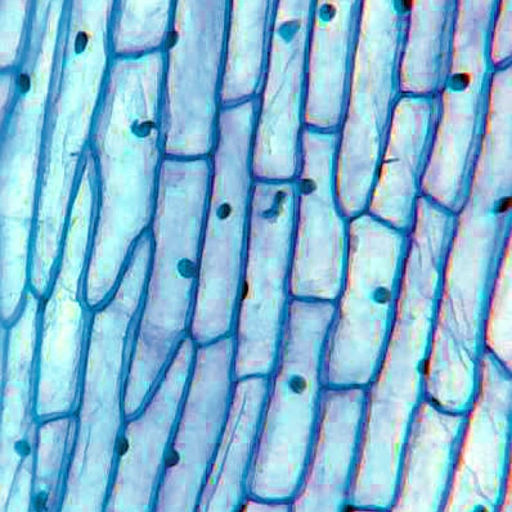
Identifying Parts of an Onion Cell
After preparing an onion cell slide and using proper microscope magnification techniques, you’re set to identify the different parts of an onion cell. Here’s a step-by-step guide to what to look for:
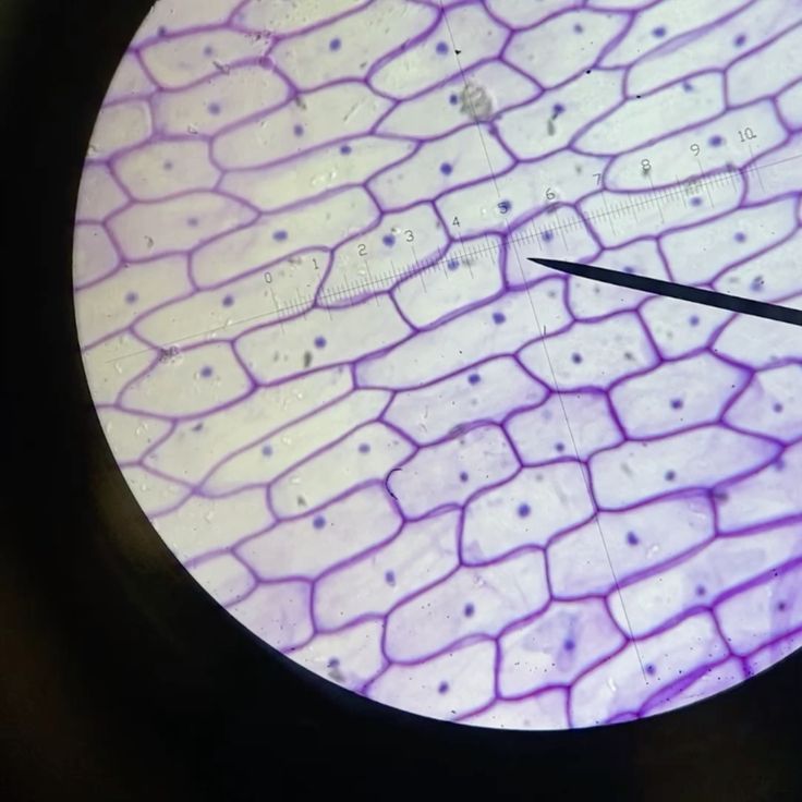
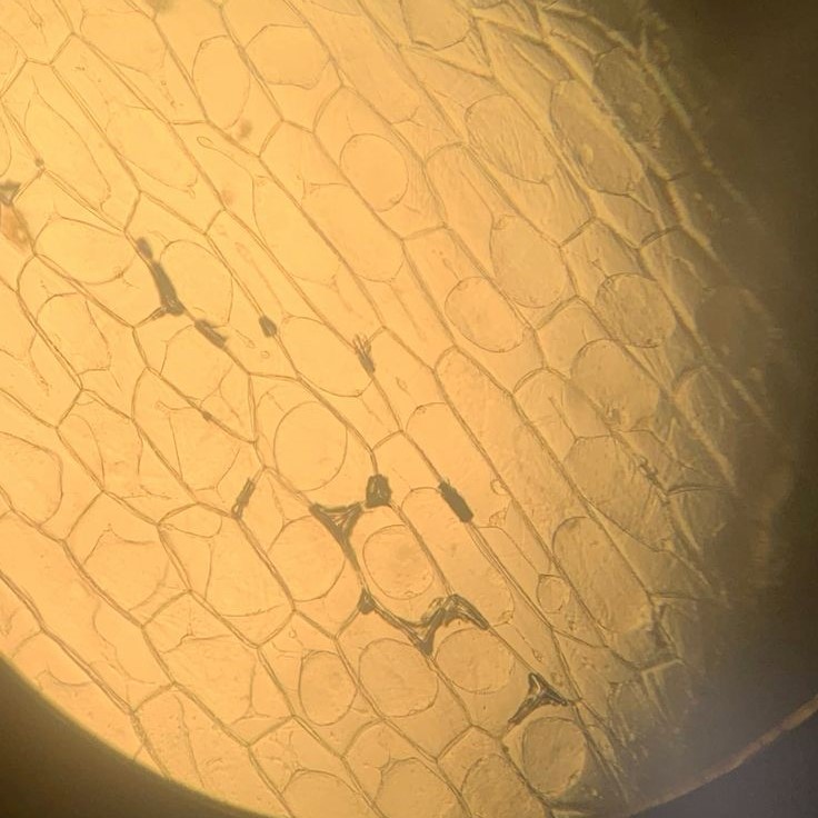
- Look for the Cell Wall: The cell wall is the outermost layer. It is rigid and gives the cell its shape.
- Identify the Cell Membrane: Just within the cell wall, find the cell membrane. It’s a thin layer that controls what enters and exits the cell.
- Spot the Cytoplasm: Inside these layers lies the cytoplasm. It’s the fluid that fills the cell. Organelles float in this jelly-like substance.
- Find the Nucleus: Look for a round or oval structure. This is the nucleus, the cell’s control center.
- Notice Mitochondria: These structures may be harder to see. They’re the cell’s powerhouses, generating energy.
- Observe Vacuoles: Visible as clear or slightly darker spots, vacuoles store nutrients and waste.
Each part has a vital role, ensuring the cell’s health and function. Use the keywords ‘onion cell under microscope’ to deepen your knowledge and find detailed images that help with identification. When you know what to look for, each structure stands out, telling a piece of the story of life at a cellular level.
The Role of the Cell Wall and Membrane
In any exploration of an onion cell under a microscope, understanding the role of the cell wall and membrane is key. Each part plays a critical job in cell life.
The cell wall acts as a strong barrier. It keeps the cell’s shape and guards against damage. Like a suit of armor, it provides structure. Made of cellulose, it’s both tough and flexible. Without it, the cell could not keep its form.
The cell membrane, on the other hand, is the gatekeeper. It’s the layer right inside the cell wall. This membrane chooses what can enter and exit the cell. Thus, it keeps the cell’s internal environment stable. This selection is crucial for cell health. Because of this control, the cell can stay alive and work well.
Both the wall and membrane are visible when viewing an onion cell under microscope. They outline the cell’s limits. When stained, these parts become even clearer. They appear as defined edges around the filled cytoplasm.
In summary, the cell wall offers protection and form. The cell membrane manages what comes in and out. These roles are essential for maintaining the cell’s integrity and function. Together, they form the first line of defense and regulation for the cell’s inner workings.
Understanding Cytoplasm and Organelles
When peering at an onion cell under a microscope, the cytoplasm is immediately apparent. This clear, jelly-like substance fills most of the cell. It acts as a cushion for the organelles, each carrying out its unique task.
Organelles are like mini-factories, each with a special job. For example, the nucleus holds the DNA, which gives instructions for everything the cell does. Mitochondria are the power units. They turn nutrients into energy the cell can use. Vacuoles act as storage bins. They keep vital materials until the cell needs them.
Other organelles include ribosomes and the endoplasmic reticulum. Ribosomes make proteins. The endoplasmic reticulum transports them through the cell. Think of it as the cell’s highway system.
Each organelle is essential for the life of the cell. Without them, the cell could not function. The cytoplasm allows them to float and move where they are needed. It is the stage where the drama of life unfolds in tiny acts.
When observing an onion cell under microscope, look for these parts. You can see them doing their jobs, keeping the cell alive and busy. It’s a complex dance of life, happening right before your eyes.
Observing Nucleus and Nucleolus Behavior
Within the bustling environment of an onion cell under microscope, the nucleus stands out. It’s often round and more distinct than other organelles. Inside this control center, the nucleolus takes a central role. The nucleolus is smaller and within the nucleus. It’s crucial for making ribosomes, key to protein production.
As you observe, you’ll notice the nucleus is not always central in the cell. It shifts position based on the cell’s activities. If the cell is dividing, the nucleus becomes quite active. You might observe changes in its shape or size. This activity is part of the cell cycle, especially during mitosis.
The nucleolus behavior is also telling. A prominent nucleolus often means the cell is actively making proteins. If you see many nucleoli or large ones, it’s a sign of high activity.
By focusing on the nucleus and nucleolus, you get to glimpse the cell’s genetic command post. It’s where the blueprints for life are stored and read. Here, you can really appreciate the complexity of cellular activities. Watching these parts work, you’re peeking at the fundamentals of life itself.
Mitosis in Onion Cells
When studying an onion cell under microscope, attention often turns to mitosis. Mitosis is the process by which a single cell divides to create two identical daughter cells, and it’s a fundamental mechanism for growth and repair in all living organisms. Observing mitosis in onion cells under a microscope provides a clear view of the cell cycle in action due to the large size and transparent nature of these plant cells. Here are the phases you’ll witness:
- Prophase: At this initial stage, the chromatin condenses into visible chromosomes. The nuclear membrane starts to disappear.
- Metaphase: Chromosomes line up at the cell’s center. Spindle fibers attach to each chromosome’s centromere, preparing for division.
- Anaphase: The spindle fibers begin to shorten, pulling apart sister chromatids to opposite ends of the cell.
- Telophase: Chromatids reach the poles. Nuclear membranes form around the two sets of chromosomes, now at each end.
- Cytokinesis: Finally, the cytoplasm divides. Two new cells emerge, each with the same genetic material as the original cell.
Watching the phases of mitosis provides insights into the intricacies of cellular reproduction. The events may occur rapidly, so patience and attention to detail are necessary when observing onion cells under a microscope. Each phase contributes to the life cycle of the cell, ensuring that each daughter cell is properly equipped to survive and function.
Staining Techniques for Enhanced Visibility
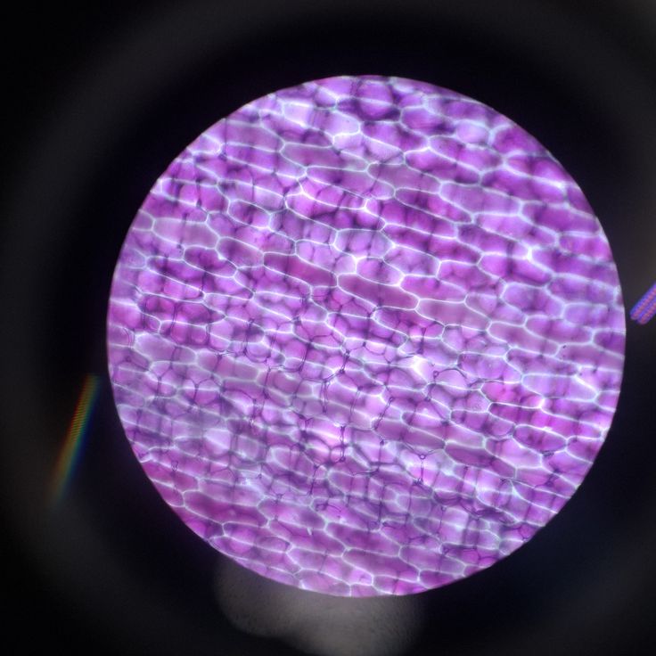
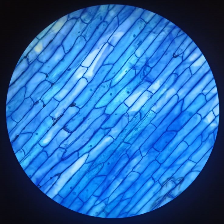
While observing an onion cell under microscope, staining techniques greatly enhance cell visibility. These techniques add color to different cell parts, making structures clearer. Let’s walk through common staining methods used for onion cells:
- Iodine Solution: This is the most common stain for plant cells. It colors the starch present in the cell, typically turning the nucleus a dark color. The contrast makes the cell’s structure stand out.
- Methylene Blue: This stain can color the cell wall and membrane, which helps differentiate these parts from the rest of the cell.
- Acetocarmine: Ideal for viewing chromosomes during mitosis. It binds to DNA and is useful for observing genetic material during cell division.
Prior to staining, ensure the onion slice is very thin. Apply just one drop of stain to prevent overwhelming the sample. Place a cover slip over it gently to avoid air bubbles or excess liquid.
Use these stains to view distinct features within the onion cell under microscope. Each stain has a unique role, highlighting various parts of the cell in different colors. This enhanced contrast aids in identifying and understanding the cell’s intricate details.