Introduction to Microscopy
When we ask, ‘what is a microscope?’ we refer to a tool that reveals the unseen. Microscopes let us explore the world beyond our naked eyes. These instruments magnify tiny objects, making them visible and measurable. Microscopy, therefore, is the science of using microscopes to study materials at the microscopic scale.
Microscopes vary in type and complexity, but they all share a common purpose. They enhance small details to better understand materials, organisms, and their structures. From school labs to advanced research facilities, microscopes are crucial for scientific discovery.
In this guide, we delve into what makes a microscope work. We will highlight its history, how it evolved, and its various types. You’ll learn about the critical parts of a microscope and their functions. We’ll also cover the concepts of magnification and resolution. Lastly, we explore the impact of microscopes in different fields and look at recent technological advancements.
Understanding how a microscope operates can open a new world of detail and precision. This knowledge can expand your curiosity and sharpen your research skills. Whether you’re a student, scientist, or hobbyist, grasping the basics of microscopy is the first step on a journey into the minuscule wonders of our world.
Historical Evolution of the Microscope
The journey of the microscope begins in the late 16th century. Two Dutch eyeglass makers, Zaccharias Janssen and his father Hans, started the evolution. They discovered that by placing lenses at each end of a tube, objects appeared larger.
By the 17th century, Galileo improved on this design, focusing on adjustable lenses. This led to the compound microscope. A pivotal moment came when Antonie van Leeuwenhoek made lenses with great magnification. His microscopes revealed bacteria, free-living and parasitic microscopic protists, sperm cells, and blood flow in capillaries.
Throughout the 19th century, optical improvements continued. The achromatic lens came into existence, reducing the problem of chromatic aberrations. By the late 19th century, Ernst Abbe formulated the theory of microscope resolution. This changed how we understood the limits of magnification.
The 20th century witnessed the emergence of new types. Electron microscopes came onto the scene, using electron beams to magnify objects. This was later followed by the invention of the scanning probe microscope.
Each evolution of the microscope built upon the last. Today, we benefit from centuries of innovation. Thanks to these advances, ‘what is a microscope’ continues to be a question with ever-expanding answers.
Types of Microscopes
When exploring ‘what is a microscope,’ one must understand the different types available. Each type has unique features and applications. Let’s examine the primary types of microscopes used in various fields.
Optical Microscopes
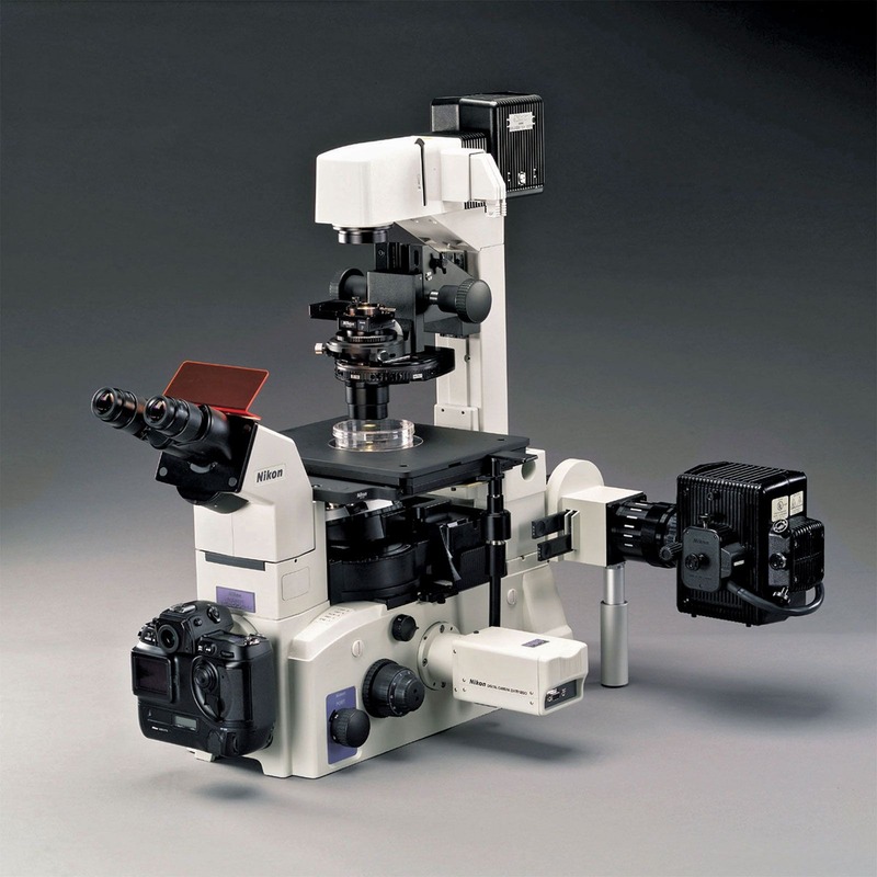
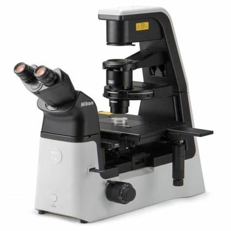
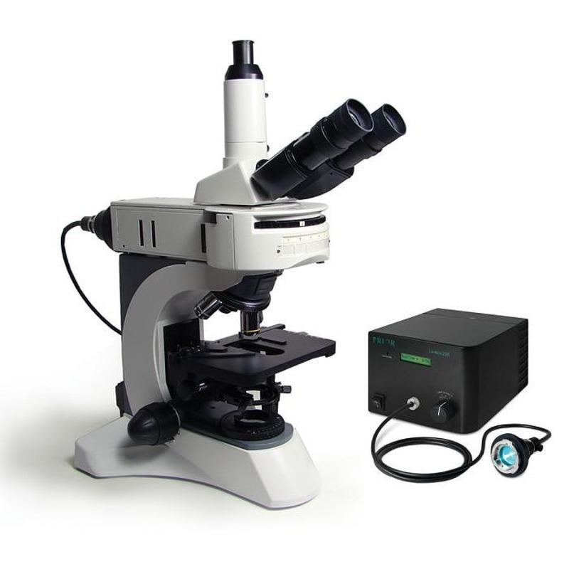
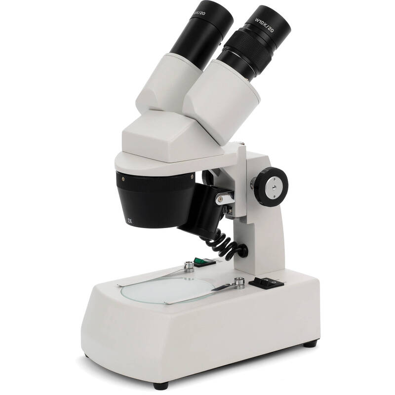
Optical microscopes, also called light microscopes, are the most common type. They use light to illuminate the sample. Optical microscopes include simple models with just one lens and compound microscopes with multiple lenses. Compound microscopes are standard in schools and research labs.
These microscopes have a range of magnifications, typically up to 1000x. They are ideal for examining cells, microorganisms, and tissues. Specialized versions, like fluorescence microscopes, can even highlight specific parts of a specimen using dyes.
Electron Microscopes
Electron microscopes offer a deeper look into the microscopic world. Instead of light, they use a beam of electrons. This allows them to achieve much higher magnifications and resolutions, sometimes over a million times.
There are two main types of electron microscopes: the transmission electron microscope (TEM) and the scanning electron microscope (SEM). TEMs provide detailed images of the internal structure of cells. SEMs, on the other hand, give a realistic 3D view of the surface of specimens.
Scanning Probe Microscopes
Scanning probe microscopes stand out by not using light or electrons. They scan the specimen with a physical probe. This technique can generate images of surfaces at the atomic level.
Atomic force microscopes (AFM) and scanning tunneling microscopes (STM) are examples of scanning probe microscopes. They are pivotal in fields like nanotechnology and materials science.
Key Components and Their Functions
To grasp ‘what is a microscope,’ one must know its key parts and their roles. Microscopes, though varied in type, contain several core components crucial for their operation.
Lenses and Objectives
Lenses are the heart of a microscope. They gather and focus light to create an image. The objective lens is vital. Placed close to the specimen, it has high magnification power. Compound microscopes usually have several objective lenses of different strengths. They let users switch between magnifications to view the sample in more detail.
The Stage and Specimen Holders
The stage is the platform where you place the sample. It holds the specimen securely and allows precise movement. Specimen holders, like slides and clips, keep samples in place during observation. They are vital for accurate focus and image clarity.
Illumination Systems
Illumination systems in microscopes shine light on the specimen. The light source might be a mirror reflecting ambient light in simple microscopes. More advanced models use built-in lamps. Proper illumination is key to a clear image. It can be adjusted to enhance contrast and details of the specimen.
Understanding Magnification and Resolution
Understanding magnification and resolution is crucial when exploring ‘what is a microscope’. Magnification refers to how much larger the microscope makes an object appear. Resolution, on the other hand, describes the ability of a microscope to distinguish between two points that are close together. Both are fundamental concepts in microscopy, affecting the quality of the images produced.
Magnification
The magnification of a microscope is the ratio of the observed size of a specimen to its actual size. Optical microscopes commonly have magnifications ranging up to 1000 times the original size of the specimen. This allows detailed observation of microscopic structures such as cells and bacteria, otherwise invisible to the naked eye.
The level of magnification is achieved through the use of lenses, primarily the objective lens and often an eyepiece lens that further enlarges the image. Magnification can reveal fascinating details, but it has limits. Beyond a certain point, increasing magnification doesn’t improve the image, because resolution becomes the limiting factor.
Resolution
Resolution is a measure of how well a microscope can separate small structures that are close together. It’s the clarity or sharpness of the image, and it’s determined by the quality of the lenses and the wavelength of light or electrons used to form the image.
For example, electron microscopes have much higher resolution than light microscopes because they use a beam of electrons with wavelengths shorter than visible light. This enables the TEM to visualize sub-cellular structures, and the SEM to produce detailed 3D images of the specimen surface.
In summary, magnification and resolution work together to provide clear and detailed images of microscopic objects. While magnification enlarges the image, resolution ensures that the finer details remain distinguishable, allowing scientists and researchers to study the intricate structures of the microscopic world.
Practical Applications of Microscopes
Microscopes are vital tools that extend beyond academic curiosity. They serve practical purposes in various critical fields.
In Medical Research
In the realm of medical research, microscopes are indispensable. They allow scientists to observe pathogens, study disease development, and monitor treatment effects. Thanks to microscopes, we can understand cell structures, identify cancerous cells, and research genetic material. They play a role in developing vaccines and in understanding how our immune system fights illness. ‘What is a microscope?’ in this context is a weapon against disease.
In Material Sciences
Material science relies on microscopes to investigate the properties of materials. Microscopes probe the atomic structures of metals, polymers, and composites. They reveal how substances interact at a microscopic level. This helps in creating stronger materials for construction, more efficient electronics, and innovative textiles. Microscopes are crucial in pushing the boundaries of what materials can do.
In Environmental Studies
Microscopes allow environmental scientists to study pollutants, assess water quality, and understand the effects of climate change on biodiversity. By examining soil and water samples, they can detect contaminants early. This use of microscopes is vital for protecting ecosystems and human health. In environmental studies, ‘what is a microscope?’ translates to a tool for conservation and sustainability.
Advances in Microscopy Technology
The field of microscopy has not stood still. Recent leaps in technology have pushed its boundaries further. ‘What is a microscope?’ today is very different from what it was even a decade ago. Innovations in digital imaging and the creation of super-resolution techniques are revolutionizing how we view the microscopic world.
Digital Imaging and Analysis
Digital imaging has transformed microscopes into high-powered computational devices. Microscopes now capture images as digital files. Researchers can store, share, and analyze data with unprecedented ease. This facilitates collaboration across the globe. Software tools enhance images and extract valuable information. Data that would have taken days to interpret now takes only minutes.
Super-Resolution Techniques
Super-resolution microscopy breaks the limits of traditional microscopy. These techniques allow scientists to view objects even smaller than the light wavelength. They refine the image resolution beyond standard capabilities. For example, techniques like STED and PALM make it possible to look at molecular processes in live cells. This has opened new doors in biological and medical research.
Care and Maintenance of Microscopes
Proper care and maintenance are key to keeping any microscope in top condition. They ensure that the device operates at its best, providing clear and accurate images. Whether you’re a student, researcher, or hobbyist, knowing how to care for your microscope can greatly extend its lifespan and performance. Regular attention to maintenance prevents common issues and preserves the precision and quality required for complex analyses. Here are essential tips for the care and maintenance of microscopes.
Handle with Care
Microscopes are delicate instruments. Always carry them with a firm grip and proper support. Avoid jarring movements or impacts that can misalign components.
Clean Regularly
Dust and oils can blur lenses, affecting magnification and resolution. Use a soft, lint-free cloth and appropriate cleaning solutions to wipe optical surfaces gently.
Check Alignment
Ensure the lenses and illumination systems are properly aligned. Misalignment can cause poor image quality and strain during observation.
Store Carefully
When not in use, cover the microscope with a dust cover and store in a dry, cool place. Avoid places with temperature swings and high humidity.
Perform Routine Inspections
Look for loose screws, misaligned parts, or signs of wear. Address problems early to prevent further damage. Professional servicing may be necessary for complex issues.
Do not attempt to fix mechanical or electrical problems unless you are trained. Contact a professional to maintain the integrity of the microscope.
By following these steps, you can keep your microscope in excellent working order, ensuring it continues to reveal the wonders of the microscopic world.


