Introduction to Paramecium
Paramecium, a tiny organism, offers a window into the unseen world under a microscope. These single-celled wonders exhibit complex behaviors that captivate scientists and hobbyists alike. When one observes a paramecium under a microscope, it unlocks a vibrant microcosm of life.
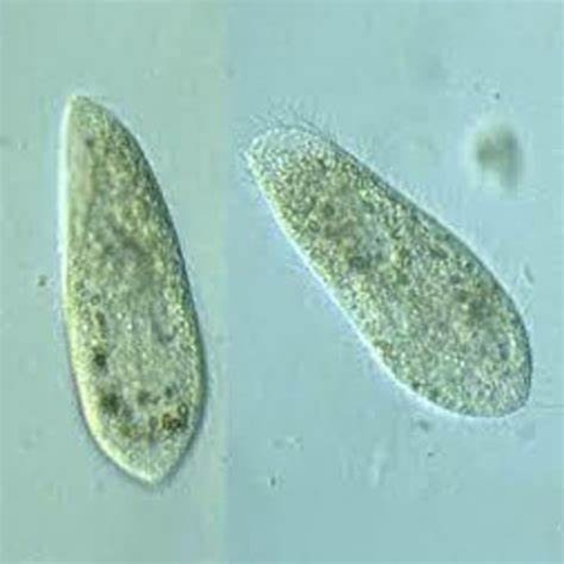
Often found in fresh water, paramecium looks pear-shaped and moves with purpose. This shape is unique and helps it in various ways. Under a microscope, you can see its tiny hairs, called cilia, beating rhythmically. These cilia are essential for movement and feeding, which one can study in detail. By examining a paramecium under microscope, curious minds can learn much about basic life functions.
Skilled at adapting, paramecium thrives in a range of aquatic environments. This makes them easy to find and study. Amateurs and experts both enjoy observing these creatures. The paramecium illustrates fundamentals of biology like locomotion, response to stimuli, and even reproduction.
Anyone exploring the micro-world can start with paramecium. They are often a first glimpse into microscopic study due to their abundance and ease of observation. Whether in a lab or a classroom, these microorganisms provide invaluable insights into the complexities of life on a scale that’s often overlooked.
To study paramecium under microscope is to begin a journey into the intricacies of life at the cellular level. It’s a journey full of discovery and wonder, and it starts with simply peering into the lens of a microscope.
The Microscopic Structure of Paramecium
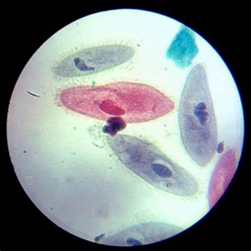
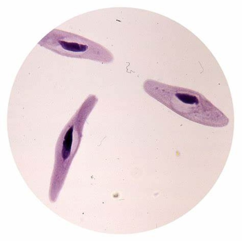
To comprehend a paramecium under a microscope, one must recognize its distinct microscopic structure. This single-celled organism is truly a marvel, boasting complex internal and external features that facilitate its survival in water. A deeper look into its microscopic structure reveals specialized components functioning in harmony.
The Cilia and Its Functions
The cilia, tiny hair-like projections that cover the paramecium’s body, are one of its most notable features. These cilia beat in coordinated waves, propelling the paramecium forward in its aquatic environment. Their rhythmic motions also create water currents. These currents guide food particles towards the paramecium’s oral groove. The cilia thus serve a dual purpose. They enable movement and contribute significantly to the creature’s feeding process.
Studies have shown that the cilia of paramecium are also sensitive to changes in the environment. They can alter their beat patterns to navigate around obstacles or towards a food source. The presence of calcium ions influence these changes, indicating a complex response system at a cellular level.
Oral Groove and Feeding Mechanisms
At the center of the paramecium’s feeding process lies its oral groove, a funnel-like structure. The oral groove channels food particles trapped by the ciliary currents into the paramecium’s mouth. As these particles accumulate, they form food vacuoles. These food vacuoles are essential. They break down the nutrients through a process called digestion.
This streamlined feeding mechanism shows the efficiency of paramecium. Despite its simplicity, the organism exhibits an advanced level of autonomy in securing sustenance. What appears to be simple structures under a microscope, the cilia, and the oral groove, are indeed critical to the paramecium’s survival.
Observing Paramecium: Preparation and Microscopy Techniques
To study paramecium under microscope, proper preparation is key. This section will guide you through collecting samples and setting up your microscope for optimal viewing.
Sample Collection
Finding paramecium starts with collecting samples from their natural habitat. Here’s how you can do it:
- Scout for stagnant or slow-moving fresh water, like ponds or lakes.
- Use a clean jar to scoop water near the surface.
- Increase your chances by collecting near plants, as paramecium feed on bacteria found there.
- Let the sample settle. Paramecium will gather at the bottom.
- Use a dropper to collect paramecium from the settled part of your sample.
Microscope Settings for Paramecium Viewing
Once you have your sample, setting your microscope correctly will help you observe paramecium in detail:
- Start with a lower magnification to find them easily.
- Once located, switch to a higher magnification for a closer look.
- Adjust the diaphragm for proper light. Paramecium are transparent and need back-lighting.
- Focus slowly, and use fine adjustments to keep the paramecium in view.
- Move the slide gently to follow their movement as they swim.
With these steps, you can observe the fascinating world of paramecium up close.
Paramecium Behavior and Movement
To truly appreciate paramecium under a microscope, one must observe their dynamic behavior and movement. Their responsiveness to the environment showcases their survival instincts. Paramecium’s movements are not random but rather highly coordinated responses to stimuli. They can avoid obstacles, find food, and escape predators. This intelligence, albeit on a cellular level, is fascinating to witness.
Response to Stimuli
Paramecium are remarkably sensitive to external stimuli in their aquatic world. They react to chemical signals, changes in light, and even touch. For instance, if they encounter a barrier, they will reverse the beat of their cilia. This action allows them to change direction seamlessly. Watching paramecium under microscope gives a clear view of their adaptations. Their responses help them thrive in a world full of variables.
Locomotion Patterns
Observing paramecium move is a captivating experience. They glide through water with a smooth, forward motion known as ‘ciliary beating’. They can also rotate around their axis as they advance. This twisting movement helps paramecium explore their environment thoroughly. The efficiency of their locomotion patterns is a testament to their evolution. It ensures their success as they navigate the microscopic world they inhabit.
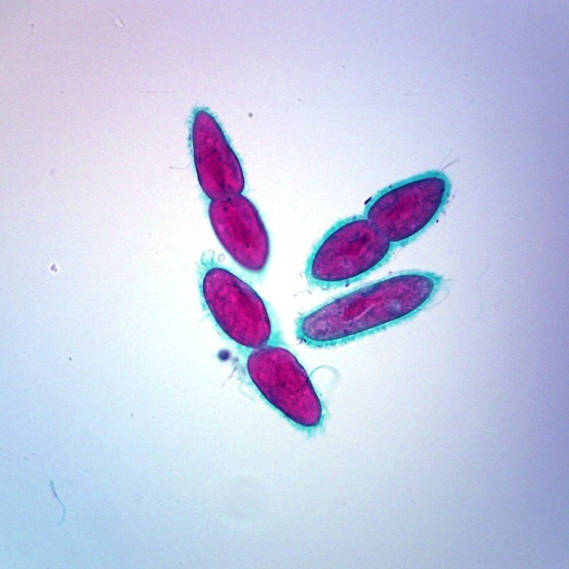
Reproduction and Lifecycle of Paramecium
The study of paramecium under a microscope gives valuable insight into their fascinating reproduction and lifecycle. These single-celled creatures have unique ways to ensure their species thrives in aquatic environments. By observing their reproduction, we gain a deeper understanding of cellular processes.
Asexual Reproduction Process
Paramecium primarily reproduce asexually through a process known as binary fission. This method is simple and efficient, allowing for rapid population growth. During binary fission, the paramecium’s nucleus divides first, followed by the division of its cytoplasm. This creates two genetically identical offspring, each inheriting half of the parent’s body and organelles.
Asexual reproduction in paramecium is a continuous cycle. It happens when conditions are good, like when there’s plenty of food and the water is at the right temperature. It’s a straightforward way for paramecium to multiply and maintain their numbers in their habitat.
Sexual Reproduction: Conjugation
Paramecium also have a form of sexual reproduction called conjugation. Conjugation is more complex than asexual reproduction and usually occurs when conditions become stressful, such as when nutrients are scarce.
During conjugation, two paramecium attach side by side and form a cytoplasmic bridge between them. Through this bridge, they exchange genetic material. This exchange results in greater genetic diversity within the paramecium population. This diversity is critical for adapting to changes in their environment and ensures the resilience of their species.
Both asexual and sexual reproduction modes serve different purposes in the lifecycle of a paramecium. Asexual reproduction allows for quick population growth, while sexual reproduction through conjugation contributes to genetic variation and survival under harsh conditions.
By watching paramecium under the microscope during these processes, we can observe the remarkable ways in which these microorganisms proliferate and adapt. Their lifecycle is a microcosm of survival and adaptation at the cellular level.
Paramecium as a Model Organism in Research
Paramecium have become keystones in the world of scientific research. Their simple structure and rapid lifecycle make them perfect for studying cell biology. They show us how cells grow, divide, and respond to genetic changes along with environmental signals. This research has wider implications for understanding human diseases as well.
Studying Cell Biology and Genetics
In genetics, paramecium offer unique insights. Their asexual and sexual reproduction methods allow for various genetic studies under a microscope. We can induce mutations, track gene expression. We can even observe the effects of genetic recombination. These observations are valuable for understanding DNA repair mechanisms.
Understanding Ecosystems and Protist Diversity
Paramecium play a role in ecosystem dynamics due to their position in the food web. Studying them under a microscope helps us grasp how protists contribute to nutrient cycles. It also shows us their response to ecological changes. By researching paramecium diversity, we understand how single-celled organisms evolve and interact with each other.
Paramecium in the Classroom
Paramecium under microscope bring biology to life for students. These organisms are simple to study. They demonstrate a range of biological processes firsthand. Classes learning about cell structure, genetics, or ecosystems can benefit greatly. Paramecium provide a practical example of the tiny worlds existing in a droplet of water.
Teachers often include them in their curriculum. This is because paramecium are easy to find and observe. They offer a clear and immediate experience of the microscopic world. With paramecium, students can see biology, not just read about it. This hands-on approach aids understanding and retention of complex concepts.
Engaging Students with Live Specimens
Live paramecium specimens grip student attention. Observing these creatures fosters curiosity and excitement. It’s one thing to discuss cilia or asexual reproduction. It’s quite another to see these features and processes in action.
Students can gather samples from local ponds. They then prepare slides to study paramecium under a microscope. This whole process involves them actively. It also strengthens their observation skills. It is an effective way to make learning dynamic and interactive.
Microscopy Skills and Science Education
Learning to use a microscope is a vital skill in science. Studying paramecium under microscope introduces this tool to students. They learn to adjust magnifications, focus on specimens, and track moving organisms.
These skills are not just for observing paramecium. They apply to a wide range of scientific inquiries. Students become familiar with equipment they may use in advanced studies or careers. Moreover, microscopes foster a meticulous approach to scientific experiments. Allowing students to develop precision and an eye for detail early on.
Conclusion: The Significance of Paramecium Study
The study of paramecium under microscope is full of insights. It shows us life at a cellular scale. This tiny organism helps us understand bigger ideas in biology. Paramecium are more than just a microbe; they are a tool for learning.
Paramecium allow us to see how life functions. They show us how a single cell can eat, move, and reproduce. They thrive in fresh water. These actions seem simple, yet they are complex. They help us grasp the basics of life.
Scientists use paramecium to research cell biology. They study genetics, diseases, and ecology with them. The simplicity of paramecium makes them ideal for such studies. They are easy to find, observe, and understand. Yet, they can teach us complex biology lessons.
In classrooms, paramecium bring science to life. Students watch them move and eat. They see how paramecium respond to light and touch. These observations make scientific concepts real. They help students learn and remember.
Paramecium illustrate big biological principles. They show us how life works on a tiny scale. Through them, we learn about the diversity of organisms. We see food chains and ecosystems in action. Paramecium are more than just an object under a lens. They are a window into the vast world of biology.
By studying paramecium, we get a snapshot of life’s complexity. These microorganisms have a lot to teach us. Their study is a foundation for both novices and experts in biology. They highlight the beauty and intricacy of life in a drop of water.


