Introduction to Grass Anatomy
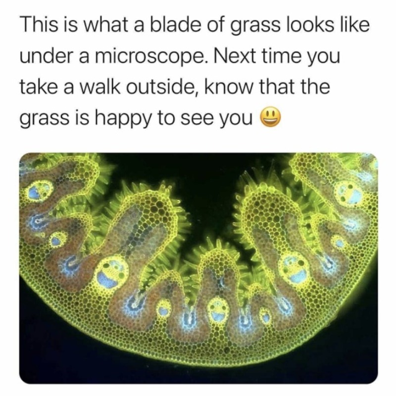
Grass, a common sight in meadows and lawns, has a complex anatomy. When we delve into ‘grass under microscope’, we uncover a whole new dimension of these ubiquitous plants. Knowing the basics of grass anatomy is key for microscopic exploration. The anatomy includes roots, stems, leaves, flowers, and seeds. Each part has unique cells and structures.
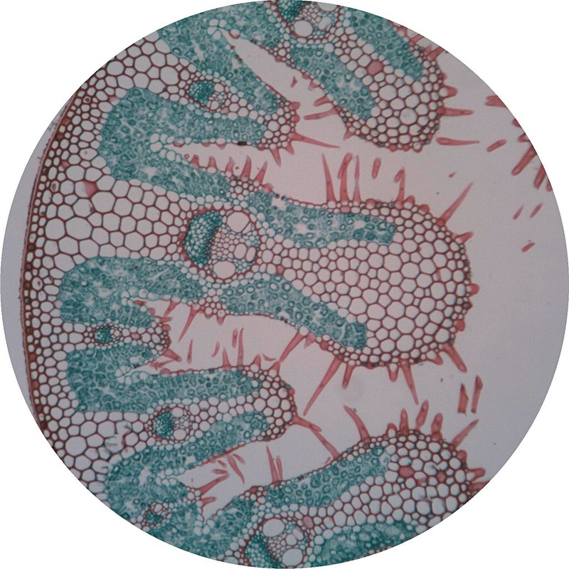
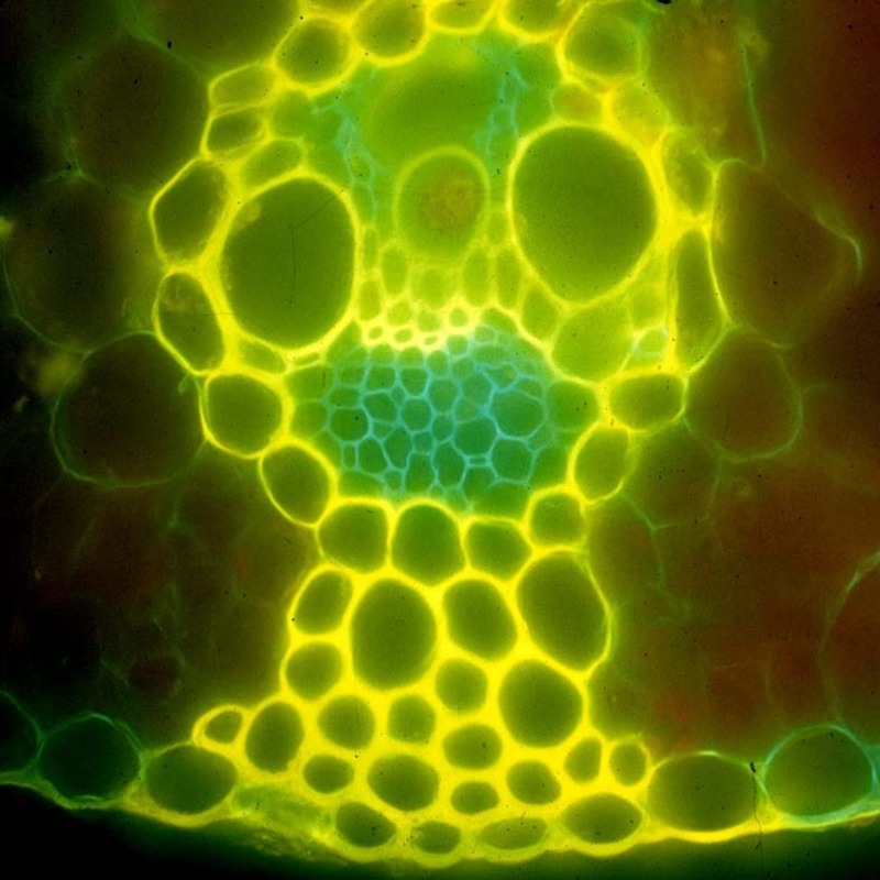
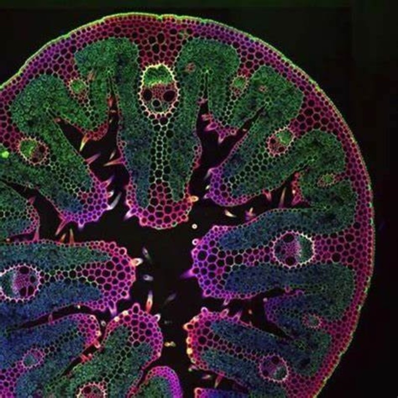
Roots anchor the grass to the soil and absorb nutrients. They show a network of root hairs under the microscope. Stems, or culms, offer support and house the vascular system that transports water and nutrients. Under high magnification, the arrangement of vascular bundles becomes evident.
Grass leaves, often the most visible part, consist of a blade and sheath. The blade is what we commonly think of as the leaf, while the sheath wraps around the stem. Microscopically, one can see the parallel veins that characterize grass leaves.
Flowers of grasses are usually less showy than other plants. Yet, they are fascinating under a microscope with intricate parts like anthers and stigma. Seeds complete the life cycle and have protective coatings and embryonic tissue, which are all observable in microscopic detail.
To conclude, ‘grass under microscope’, reveals an intricate world. From cells that capture sunlight to those that fend off pests, every component plays a vital role. Next, we’ll go over how to prepare grass samples for microscopic investigation to peer into this hidden realm.
Preparing Grass Samples for Microscopic Examination
Examining grass under a microscope begins with correct sample preparation. Here is a step-by-step guide to get your grass samples ready for microscopic observation:
Collecting Your Sample
First, choose healthy grass from an area free of pesticides. Ensure it represents the part of grass you wish to study. It could be the root, stem, leaf, flower, or seed.
Cleaning the Sample
Once collected, gently wash the grass with water. This removes any dirt that could interfere with your view. Use tweezers to handle the grass and avoid contamination.
Slicing the Sample
Thin slices of grass will transmit light better. Use a sharp blade to cut thin sections. This makes the cells and structures more visible under the microscope.
Staining the Sample
Certain dyes can highlight features of the grass. Staining helps differentiate various cell types. Basic stains like iodine can be used for simple examinations.
Mounting on Slides
Place your prepared sample on a glass slide. Use a cover slip on top to protect it. This also flattens the grass for better viewing.
Adjusting Microscope Settings
Before observing, adjust the microscope’s light and focus. This ensures clarity and detail in your images. Use a low power objective lens to start.
Remember, ‘grass under microscope’ will display cellular structures in detail. Be gentle and patient as you prepare the samples for the most informative views. This preparation is crucial for the accurate study of grass anatomy at the microscopic level.
Tools and Techniques for Microscope Observation
Using a microscope to explore grass features calls for specific tools and methods. Here’s a rundown of essential practices:
Choosing the Right Microscope
Pick a compound microscope with high magnification abilities. It should have adjustable lenses for clear, close-up views.
Using Proper Lighting
Good lighting is crucial. Employ a microscope with a built-in light source. Adjust the intensity to reduce glare and show details.
Adjusting Magnification
Begin with a low magnification. Increase slowly to find the best view without blurring.
Focusing Techniques
Use the fine focus knob after the coarse adjustment. This sharpens the image of the grass cells.
Live vs. Prepared Samples
Decide if you’ll view living grass cells or prepared, stained specimens. Each has benefits for different observations.
By mastering these tools and methods, a viewer can reveal the world of ‘grass under microscope’ with precision and detail.
The Structure of Grass Cells Observed Under Microscope
When we peer into the world of ‘grass under microscope’, we discover the minuscule structures that form the very basis of grass plants. Here, we delve into the specifics of grassroots cells observed under the microscope.
Types of Cells in Grass
Grass cells vary, each with a function. We find epidermal cells that protect the surface. They are typically flat and form a tight layer. Next, we see elongated and sturdy sclerenchyma cells. These provide support. Parenchyma cells fill the spaces; they store food and help in healing. Lastly, we have vascular cells. They form the tubes that transport water and nutrients.
Cell Wall Structure
Under the microscope, the grass cell walls stand out. They are thick and give strength to the plant. We can observe a primary cell wall, which is flexible, and for some cells, a secondary wall that is more rigid. These walls are key to a grass blade’s ability to stand upright.
Chloroplasts in Grass Cells
Chloroplasts are vital for photosynthesis. They appear as green dots under the microscope. With their own DNA, these organelles capture sunlight and create energy for the plant.
Nucleus and Organelles
The nucleus is the command center, holding the grass cell’s DNA. This, along with organelles like mitochondria and ribosomes, is visible. They keep the cell alive and functioning, contributing to the growth and health of the grass.
The Cytoplasm’s Role
The cytoplasm is a jelly-like material. It holds everything in place and allows movement of materials. It is less defined under the microscope but just as essential as the more visible structures.
Exploring ‘grass under microscope’ provides a deeper understanding of these living organisms. We see how each cell and structure plays an essential role. Microscopes give us a fascinating glimpse into the unseen, yet vital, world of grass structure.
Common Grass Species and Their Microscopic Differences
Observing ‘grass under microscope’ can reveal many differences between species. The common types of grass include Kentucky bluegrass, Bermuda grass, and fescue. Here we discuss their microscopic characteristics.
Kentucky Bluegrass
This grass shows a unique boat-shaped tip at a microscopic level. Its cells are well-ordered. This organization provides strength and resilience.
Bermuda Grass
Under the microscope, Bermuda grass cells display more pronounced starch grains. This reflects its high energy storage, useful in drought conditions.
Fescue
Fescue grass cells tend to be more elongated. Their chloroplast arrangement is distinct and aids in efficient photosynthesis.
Each grass species has adapted to its environment. By studying ‘grass under microscope’, we see these adaptations in the intricacies of their cellular design.
The Role of Grass in the Ecosystem and Its Microscopic Interaction
Grass plays a significant role in our ecosystem. Its presence is not just about aesthetics. By looking at ‘grass under microscope’, we can understand its impact on a larger scale. Here are some key roles and interactions:
- Soil Stability: Grass roots system binds soil. This prevents erosion by wind and water. Microscopic examination of roots reveals this dense network.
- Water Regulation: The root structure helps in water absorption and retention. This is crucial for maintaining groundwater levels.
- Carbon Storage: Grass absorbs carbon dioxide for photosynthesis. On a microscale, we see cells working to trap this gas.
- Habitat for Organisms: Grass supports diverse microorganisms. Many live among roots and leaves. Under the microscope, we can spot these tiny creatures.
- Oxygen Production: Through photosynthesis, grass releases oxygen. Microscopic views of chloroplasts show where this process happens.
- Food Source: Grasses are key in food chains. They feed numerous animals. Even at a cellular level, we can identify which parts store nutrients.
Seeing ‘grass under microscope’ helps us appreciate not just their beauty, but also their critical ecological functions. Every cell contributes to these roles, sustaining life around it.
Potential Diseases and Pests in Grass Detected with Microscopy
Microscopy is a powerful tool for detecting grass diseases and pests. Tiny invaders that hurt grass health often hide from the naked eye. Microscopes bring these small threats into view. Here is a look at common issues found using microscopy.
Fungal Infections
Many grass diseases come from fungi. These include rust, blight, and dollar spot. Under the microscope, you can see fungal spores that infect grass blades. These spores may appear as spots or web-like structures on the cells.
Bacterial Diseases
Bacterial infections cause wilt and rot in grass. Through a microscope, you can spot bacteria as tiny rods or spheres. These bacteria can invade and damage grass cells, leading to brown spots or slimy areas.
Viral Pathogens
Viruses affect grass too. They are tricky to spot, even with a microscope. Infected cells may show changes in structure or color. Viruses can stunt grass growth or cause unusual patterns on leaves.
Insect Infestations
Small insects, like chinch bugs and mites, feast on grass. A close-up look reveals these bugs or their larvae feasting on leaves and roots. Damage from these pests shows up as weakened, discolored, or dead patches in lawns.
Nematode Intruders
Nematodes are tiny worms that harm grass roots. Microscopic examination reveals their slender bodies within the root structures. These nematodes can cause yellow, stunted grass due to root damage.
By using microscopes, gardeners and professionals can spot these issues early. Early detection means better chances of saving the grass. It’s another layer of ‘grass under microscope’ revealing secrets of plant health.
Advancements in Microscopic Technologies for Plant Science
The recent advancements in microscopic technologies have revolutionized plant science. These new methods allow for greater detail and clarity when observing specimens, including ‘grass under microscope’. Here are some key developments:
High-Resolution Imaging
New microscopes offer high-resolution images. This lets scientists see finer details in grass cells. Structures like chloroplasts and cell walls, appear much clearer.
3D Imaging Techniques
3D imaging gives depth to observations. It makes understanding the complex structure of grass cells easier. It shows how parts fit and work together.
Fluorescence Microscopy
Fluorescence microscopy uses special stains that glow under certain light. It highlights specific parts of a cell. This helps to identify different cell components in grass quickly.
Electron Microscopy
Electron microscopes use beams of electrons instead of light. This allows for much greater magnification. We can see textures and layers within the grass cell walls that were once too small to view.
Digital Imaging Software
Advanced software helps to analyze microscopic images. It can measure and record changes over time. This makes studying the growth and health of grass more precise.
These advancements not only enhance our ability to view ‘grass under microscope’ but also push forward our understanding of its role in the ecosystem. With these technologies, researchers can now delve deeper into the microscopic world that is vital for the health of our planet.


