Introduction to Skeletal Muscle Tissue
Skeletal muscles are vital for movement and support in the human body. These muscles attach to bones, giving us the ability to control our limbs voluntarily. When viewed under a microscope, the complexity of skeletal muscle tissue becomes apparent. Within these muscles, fibers work together to produce force and motion. The texture of skeletal muscle under a microscope reveals distinct patterns. These patterns reflect the muscle’s intricate cellular organization.
Studying skeletal muscle under a microscope can reveal insights into how muscles contract and tire. It also helps in understanding various muscle-related diseases. Health professionals and researchers examine skeletal muscle tissue samples to diagnose conditions. This can lead to better treatment and preventative measures for muscle disorders. The analysis of these muscles at the microscopic level is a cornerstone in medical science. It bridges the gap between macroscopic understanding and cellular-level insights.
To appreciate what makes up skeletal muscle tissue, it is helpful to know its basic building blocks. These include fibers, myofibrils, sarcomeres, and neuromuscular junctions. Each structure has a unique role and is visible under microscopic examination. For example, myofibrils are the basic units that enable muscles to contract. The sarcomere, within myofibrils, is the actual contractile unit of the muscle. The neuromuscular junction is where the muscle fiber receives signals to initiate movement.
In the following sections, we’ll dive deeper into the microscopic anatomy of skeletal muscle. We’ll also explore how to prepare tissue samples and identify different fiber types under a microscope. Understanding the role of myofibrils and sarcomeres will also be covered. A closer look at neuromuscular junctions and common pathologies seen under the microscope will round out our exploration.
The Microscopic Anatomy of Skeletal Muscle
When the skeletal muscle is observed under a microscope, a world of intricate detail unfolds. Microscopic anatomy reveals the interaction between various components that make up a muscle fiber. Here, we delve into the elements visible at the micro-level and how they work together.
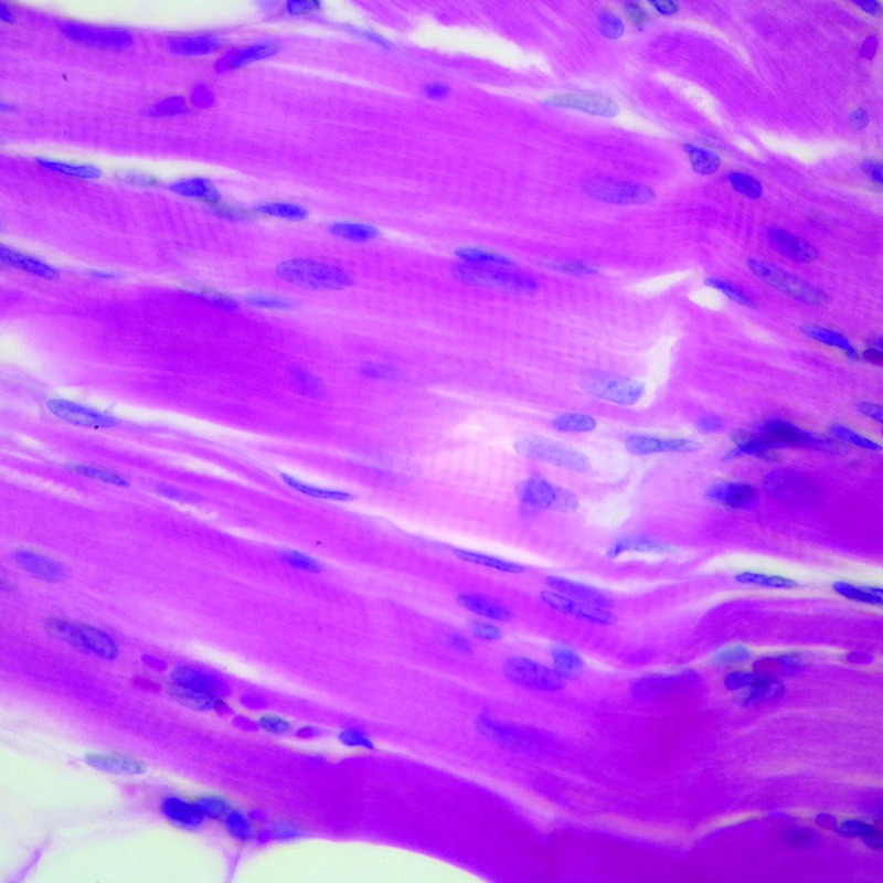
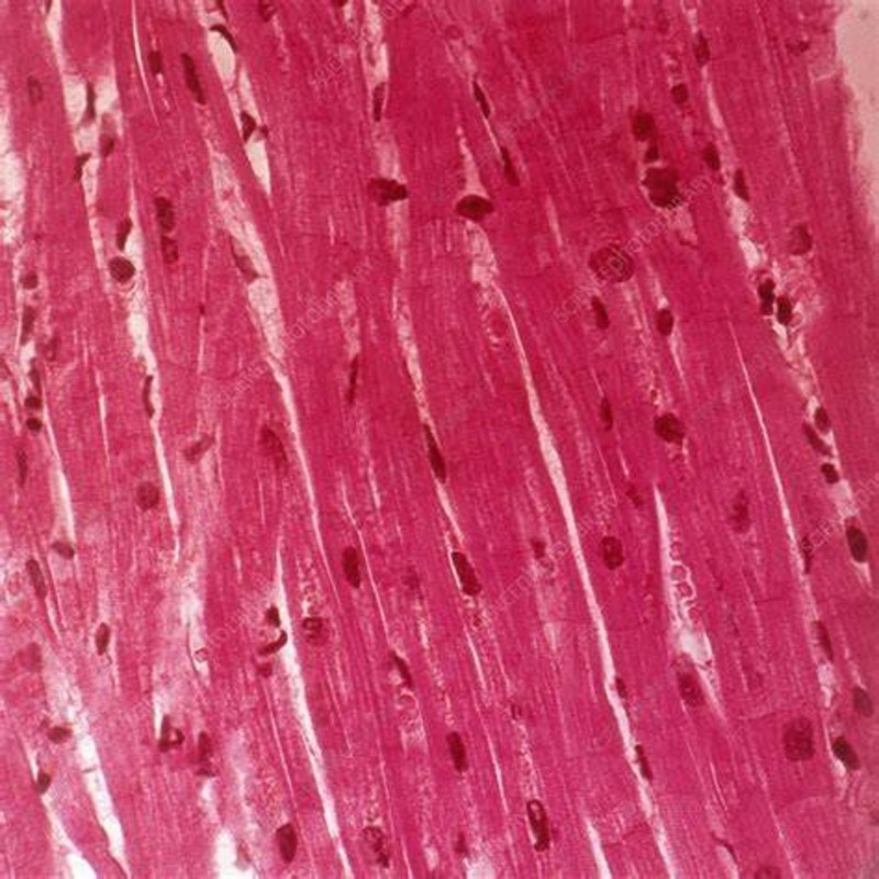
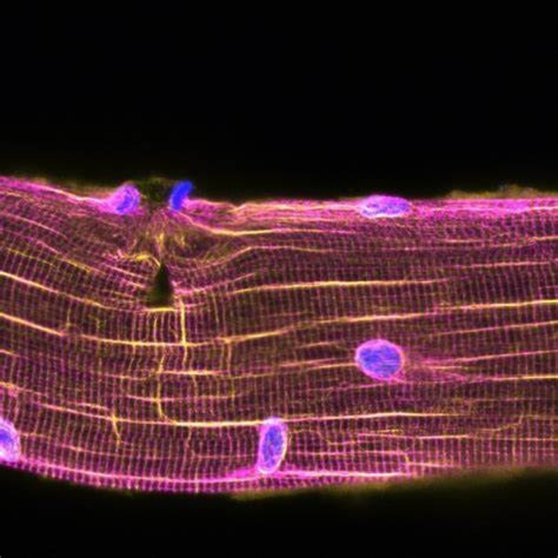
Muscle fibers are the primary structural units of skeletal muscle, appearing as long, cylindrical cells. Within these fibers, myofibrils run parallel, giving muscles their striped appearance, known as striations. Each myofibril comprises repeating sections called sarcomeres, vital for contraction.
Surrounding the muscle fibers is the endomysium, a layer of connective tissue that provides support and carries nerves and blood vessels. Adjacent muscle fibers bundle together, surrounded by another connective tissue layer called the perimysium, forming a fascicle. Many fascicles group together, wrapped in the epimysium, to form the entire muscle.
Nuclei and mitochondria are also integral to muscle fiber’s function. Scattered along the length, nuclei manage cellular activities, whereas mitochondria produce the energy necessary for muscle contraction. Together, they maintain the health and functionality of the muscle cells.
Understanding the microscopic anatomy of skeletal muscle is essential for identifying how these muscles maintain stamina and recover from fatigue or injury. It also aids in diagnosing diseases and crafting effective therapies for muscle-related conditions. By studying skeletal muscle under a microscope, scientists and medical professionals can grasp the full complexity and beauty of these critical structures in the human body.
Preparing Muscle Tissue Samples for Microscopic Examination
Before examining skeletal muscle under a microscope, proper preparation of tissue samples is critical. This process ensures clear and accurate observations. Here are the essential steps to prepare muscle tissue for microscopic examination:
- Collection: The first step is obtaining a fresh muscle tissue sample. This is often done through a biopsy or during surgery.
- Fixation: The sample is then treated with chemicals, such as formaldehyde, to preserve it. Fixation prevents decay and maintains the tissue’s structure.
- Dehydration: After fixation, the sample undergoes dehydration. This involves removing all water content, typically using a series of alcohol washes.
- Clearing: Next, the dehydrated tissue is cleared with a substance like xylene. Clearing makes the tissue more permeable to embedding materials.
- Embedding: The sample is embedded in a medium like paraffin wax. This provides support and makes it easier to cut into thin slices.
- Sectioning: Using a microtome, the embedded tissue is sliced into thin sections. These sections should be thin enough to allow light to pass through for microscopic viewing.
- Mounting: The thin slices are then placed on a glass microscope slide. They are carefully flattened and adhered to the slide.
- Staining: Finally, the tissue is stained to highlight different structures. Common stains include hematoxylin and eosin.
Each of these steps is crucial for viewing the skeletal muscle’s microstructures effectively under the microscope. Handling the tissue gently and with precision at each stage can prevent artifacts that could distort the image. With careful preparation, the sample is ready for detailed examination, revealing the remarkable complexity of skeletal muscle tissue.
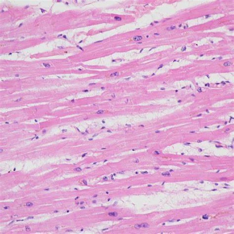
Common Staining Techniques for Muscle Observation
When we observe skeletal muscle under a microscope, staining techniques are key. They bring out details not seen in the unstained tissue. Stains add contrast to muscle microstructures, making them easier to distinguish. Here are some common staining methods used:
- Hematoxylin and Eosin (H&E): This is a widely used staining technique. Hematoxylin stains the cell nuclei blue, while eosin makes the cytoplasm and connective tissue stand out in pink and red hues.
- Trichrome Stains: These can further differentiate muscle components. Trichrome stains color muscle fibers in red or green, collagen in blue or green, and nuclei in black or red, providing a sharp contrast of muscle tissue layers.
- Immunohistochemistry (IHC): IHC targets specific proteins in muscle fibers using antibodies. This creates visual markers for researchers to identify certain muscle diseases.
- NADH-TR Staining: This technique reveals the distribution of mitochondria. It uses the NADH enzyme to create a reaction that results in a dark color in areas rich in mitochondria.
- ATPase Staining: This method differentiates muscle fibers based on their contraction speed. It adjusts the pH to react differently with various fiber types, changing their color.
Each staining technique serves a unique purpose. It highlights different features of the skeletal muscle under microscope examination. The choice of staining depends on the research focus or the specific diagnostics needed. With the help of these stains, researchers and medical professionals can observe the intricate world within muscle tissue. This insight is crucial for understanding muscle function and diagnosing diseases.
Identifying Muscle Fiber Types Under the Microscope
Identifying different muscle fiber types under a microscope is a critical process in muscle physiology. Each type of muscle fiber has unique properties that determine how a muscle functions. Generally, muscle fibers can be categorized into two main types: slow-twitch and fast-twitch fibers.
Slow-Twitch (Type I) Muscle Fibers
These fibers are designed for endurance. They contract slowly and can sustain activity for a long time without tiring. They appear darker under the microscope due to a richer blood supply and more myoglobin, which stores oxygen.
Fast-Twitch (Type II) Muscle Fibers
Fast-twitch fibers are all about speed and power. They contract quickly and strongly but tire out fast. Under the microscope, they look lighter, with less myoglobin. There are further subcategories here, such as Type IIa and IIb, differing in power, speed, and endurance.
To identify these fibers, specific staining methods are employed. ATPase staining is particularly useful. It changes the color of the fibers based on their contraction speed, helping researchers distinguish between them.
Analyzing the proportion of these fiber types within a muscle can reveal its primary function. For example, a sprinter’s leg muscle would have a higher concentration of fast-twitch fibers. Conversely, a marathoner’s leg muscle would primarily consist of slow-twitch fibers.
Understanding the distribution and characteristics of muscle fibers enhances our knowledge of muscle function. It also aids in the diagnosis of muscular conditions and informs training decisions for athletes. For anyone serious about fitness or muscle health, recognizing the details of muscle fiber types under the microscope is invaluable.
Understanding the Role of Myofibrils and Sarcomeres
Myofibrils are the engines of muscle fibers. They run the length of the fiber and contain sarcomeres. These sarcomeres are the true heroes of muscle contraction. They shorten when a muscle contracts and stretch out when it relaxes. Under a microscope, the sarcomeres display a pattern of light and dark bands. The light bands, or I-bands, contain thin actin filaments. The dark bands, called A-bands, hold thick myosin filaments.
During a muscle contraction, myosin heads hook onto actin filaments. They pull them inward, which shortens the sarcomere. Each muscle fiber has thousands of sarcomeres that work together. This coordinated action allows for powerful, precise movements.
Studying skeletal muscle under microscope can reveal the health of these structures. Damage or mutation can lead to muscle weakness and disease. By understanding myofibrils and sarcomeres, scientists can develop treatments. These treatments aim at improving muscle function and aiding recovery. Myofibrils and sarcomeres are critical for both everyday activities and athletic performance. So, their study is vital for medical science and human health alike.
The Neuromuscular Junction: A Closer Look
The neuromuscular junction is a critical bridge between the nervous system and muscular response. It’s where a motor neuron transmits a signal to a muscle fiber. This prompts the muscle to contract. A careful study of the neuromuscular junction under a microscope can shed light on how these signals are communicated. It also reveals the detailed structure that supports this process.
Under high magnification, you can see the synapse. This is where the neuron meets the muscle. Neurotransmitters like acetylcholine are released here. They cross the synaptic gap and bind to receptors on the muscle cell. This binding opens ion channels, leading to muscle excitation and contraction.
By staining neuromuscular junctions, scientists can observe specific proteins. These are crucial for signal transmission. Abnormalities in the structure or function of the junction can lead to muscle diseases. For example, myasthenia gravis is one such condition. It is characterized by weakness and fatigue of voluntary muscles.
The neuromuscular junction, while small, is incredibly complex and powerful. Its health is vital for our ability to move and interact with the world. Through studying it under a microscope, we gain valuable insights into muscle function and potential treatment for related diseases.
Pathologies of Skeletal Muscle Visible Under Microscopy
Studying skeletal muscle under a microscope also involves identifying various pathologies. These are conditions that can affect muscle function and health.
Muscular Dystrophy: This group of diseases causes progressive weakness and loss of muscle mass. Under the microscope, one may observe irregular sizes of muscle fibers and increased connective tissue.
Myopathies: Characterized by muscle weakness, myopathies often show variations in fiber size, internal structure disruptions, and abnormal deposits within the fibers.
Inflammation and Infections: Conditions like myositis involve the inflammation of muscle fibers. Microscopic examination may reveal swollen fibers and infiltration by immune cells.
Denervation Atrophy: This occurs when nerve supply to a muscle is cut off. Muscle fibers may appear much smaller and are sometimes replaced by fat tissue.
Metabolic Disorders: Diseases such as mitochondrial myopathies affect energy production. They may present with abnormal mitochondrial accumulation under the microscope.
Detecting these conditions under the microscope helps in making accurate diagnoses. It aids in the creation of treatment plans to improve or manage symptoms. Skilled interpretation of the microscopic changes in muscle tissue is crucial for medical professionals involved in muscle health and research.


