What is Euglena?
Euglena is a microorganism that scientists often study under a microscope. These single-celled creatures belong to the genus Euglena. They are well-known for their ability to move and feed like animals, and to photosynthesize like plants.Explore Euglena under microscope! Learn about its structure, ecological significance, and tips for observing this fascinating organism.
The Unique Characteristics of Euglena
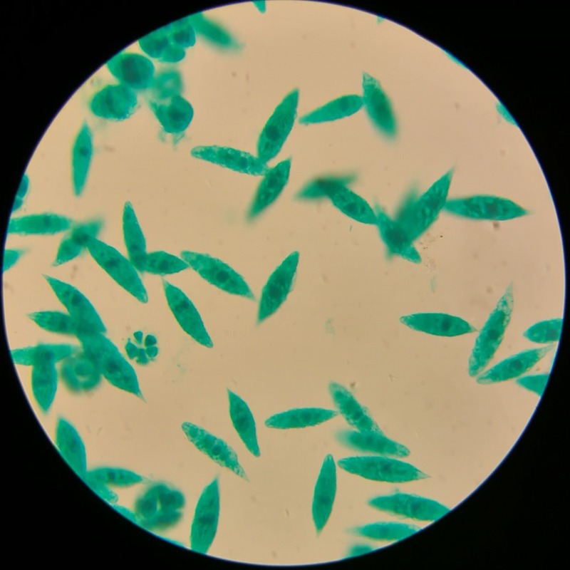
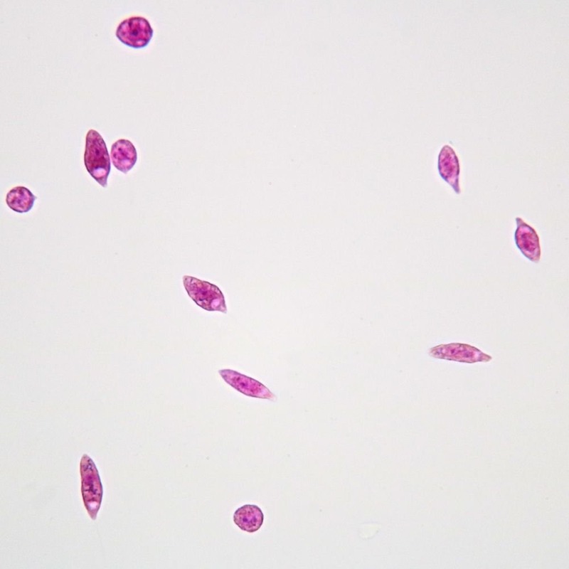
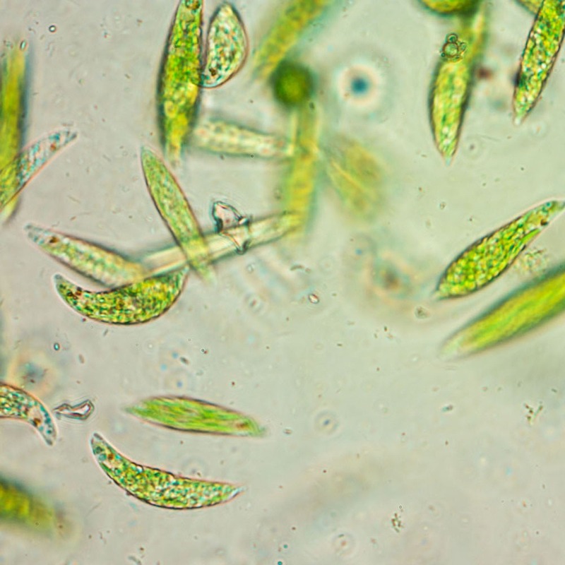
Euglena’s shape is like a spindle, and it can twist and turn actively. It has a red eye-spot that senses light, aiding in photosynthesis. Euglena’s outer layer, the pellicle, lets it change shape. With chloroplasts inside, Euglena can make its food using sunlight. It thrives in freshwaters, such as ponds and marshes.
The Role of Euglena in the Ecosystem
In ecosystems, euglena under microscope appears as both consumer and producer. It eats small particles when light is low and photosynthesizes when it’s bright. Euglena can be food for other microbes and small aquatic animals. This way, it supports higher food chain levels. Also, by photosynthesizing, Euglena helps reduce carbon dioxide levels and provides oxygen. It contributes to the balance of aquatic ecosystems.
Preparing for Euglena Observation
Before delving into the microscopic world to observe euglena under microscope, adequate preparation is essential. This involves gathering the right equipment and preparing samples in a manner that preserves the integrity of the euglena. Proper preparation is crucial to obtaining clear and informative observations.
Equipment Needed for Microscopic Examination
To observe euglena under microscope, certain pieces of equipment are indispensable. Firstly, you require a reliable microscope with variable magnification settings. Ideally, this microscope should have the capability to magnify up to 400x – allowing for a detailed view of the euglena. You’ll also need microscope slides and coverslips to hold your samples. Additionally, a pipette or dropper is necessary for transferring water samples containing euglena to your slides. Ensure all equipment is clean to avoid contamination.
Sample Collection and Preparation
Collecting euglena samples involves visiting a freshwater source, such as a pond or marsh, where euglena naturally thrive. Use a collection jar or container to scoop up some water, aiming for areas with ample sunlight exposure since euglena are often found in these conditions. Once collected, it’s important to prepare the samples carefully for examination. Place a small droplet of water on a microscope slide, then gently lay a coverslip on top, avoiding air bubbles. This preparation aids in keeping the euglena in a single layer for more straightforward observation under the microscope.
Step-by-Step Guide to Observing Euglena
Viewing euglena under microscope requires a systematic approach for best results. Follow this guide for a rewarding observation experience.
Focusing on the Microscope
Begin with the lowest magnification. Look for the green tint that indicates euglena presence. Adjust the focus slowly. Use fine adjustments to clarify the image. Moving up in magnification, refocus after each increase.
Identifying Euglena Structures
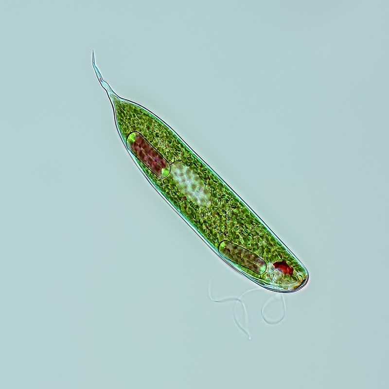
Observe the spindle shape characteristic of euglena. Locate the red eye-spot, usually near the front. Find the flagellum, a whip-like structure aiding movement. Note the green chloroplasts scattered throughout the body. Watch for euglena’s contractile vacuole, used to expel water.
Understanding Euglena Movement
Understanding how Euglena moves is crucial to observing these microorganisms effectively.
The Flagellum and Motility
Euglena propels itself with a whip-like structure called a flagellum. This tail-like appendage beats in a rhythmic manner, generating movement. With it, Euglena can navigate through water in a forward motion, often in spirals or zigzags. It’s fascinating to observe this under a microscope, where their swift swimming patterns become visible. By adjusting the microscope, viewers can see the flagellum in action, making ‘euglena under microscope’ an engaging search for scientists and enthusiasts alike.
Behavioral Responses to Environmental Stimuli
Euglena reacts to various environmental stimuli, displaying a range of behaviors. Light is a key factor. Euglena will move towards light sources, a behavior known as positive phototaxis. Conversely, it will often avoid dark areas, indicative of negative phototaxis. Temperature and chemical cues also affect Euglena’s movement. By observing these responses under a microscope, one can gain insight into how euglena interacts with its environment. This not only showcases its complexity but also underscores the importance of precise conditions for ‘euglena under microscope’ observations.
Euglena Reproduction and Lifecycle
Reproducing mostly asexually, Euglena divides by binary fission. This intriguing process ensures rapid population growth when conditions are right. To observe euglena under microscope, it is fascinating to see cell division as it occurs. Understanding this cycle is key to studying Euglena’s role in aquatic environments.
Asexual Reproduction in Euglena
Euglena reproduces asexually, splitting itself into two identical cells. In a nutrient-rich environment, Euglena thrives, repeatedly replicating through binary fission. Your microscope will reveal a Euglena elongating and its nucleus duplicating. Then, it constricts around the middle, eventually separating into newly independent organisms.
The Stages of Euglena Lifecycle
The lifecycle of Euglena consists of several stages. Initially, a mature Euglena prepares by replicating its DNA. Next, the cell elongates and the nucleus divides. Following nuclear division, the Euglena starts to pinch in two. Finally, two new Euglenas emerge, each carrying the same characteristics as the parent. They are now ready for another cycle of life. Euglena under microscope viewing can sometimes catch these stages, offering a glimpse into cellular life.
Photomicrography Tips for Euglena
Capturing images of euglena under microscope provides both a challenge and an opportunity to explore microscopic photography, or photomicrography. Here are some tips to get the best out of your euglena observations.
Lighting and Magnification Tricks
Good lighting is essential when looking at euglena under microscope. Use direct light to make euglena’s features stand out. Start with low magnification to locate euglena, then gradually increase it for detail. Finally, adjust the diaphragm to balance the light just right.
Remember to avoid overexposure. Too much light can wash out the details. Try side illumination for contrast. This highlights euglena’s unique structures. And if your microscope has it, use polarized light. It can reveal hidden details in euglena’s structure.
Capturing Clear Images of Euglena
To get clear pictures, stabilize your microscope. Any shaking can blur the image. Use a camera with a high-resolution for quality captures. If connecting your camera to the microscope, align it well. This ensures the camera sees exactly what you do.
Focusing precisely is key. Move the focus knob slowly, watch for sharp edges. Sometimes, live euglena move fast. Use a high shutter speed to freeze their motion. And for sharpness across the whole image, use a technique called focus stacking. This combines multiple pictures, each focused on different depths, into one detailed image.
To sum up, with the right tricks, observing and photographing euglena can be both fascinating and educational. Remember these tips next time you peer into the microscopic world.
Common Questions and Misconceptions
When exploring the micro world, especially with euglena under microscope, questions and misconceptions often arise. It’s important to address these to deepen understanding and appreciation for these microscopic organisms.
Differentiating Euglena from Similar Microorganisms
Identifying euglena can sometimes be tricky, as they share traits with other microorganisms. Here are some tips to help differentiate them:
- Look for the unique spindle shape that’s characteristic of Euglena.
- Spot the red eye-spot, which is not common in other microorganisms.
- Search for the flagellum, the whip-like structure enabling movement.
These features should help distinguish euglena from other similar creatures you might encounter under the microscope.
Debunking Myths About Microscopic Life
Some people think microscopic life is simple or not important. This is not true. Euglena under microscope show complex behaviors and have a big role in ecosystems. They photosynthesize like plants and move like animals. Understanding these small organisms helps us see the big picture of life on Earth.
By recognizing the complexities of euglena and similar organisms, we can dismiss common myths and gain a true appreciation for the diversity of microscopic life.