
Revolutionary Microscopes of 2025
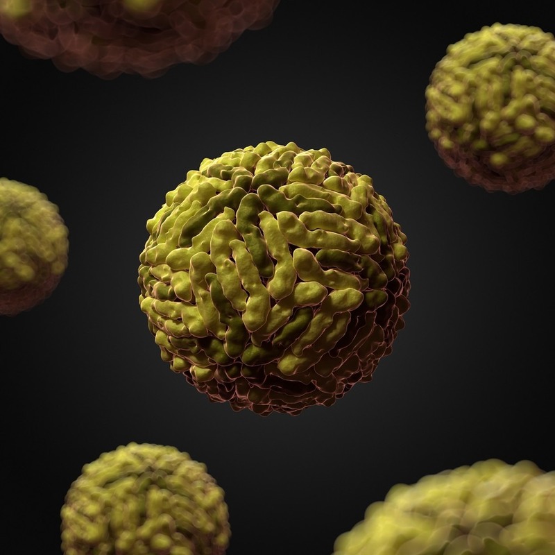
The year 2025 marks a new era in virology, thanks to innovative microscopes. These groundbreaking tools allow us to see a virus under a microscope like never before. Their enhanced capabilities are transforming our understanding of viral structures and behaviors.
Key developments in this area include high-throughput screening microscopes. They capture detailed images of viruses in real time. This lets researchers monitor viral interactions with unprecedented clarity. Another advance comes from digital imaging. It sharpens viral images further, which aids in accurate analysis.
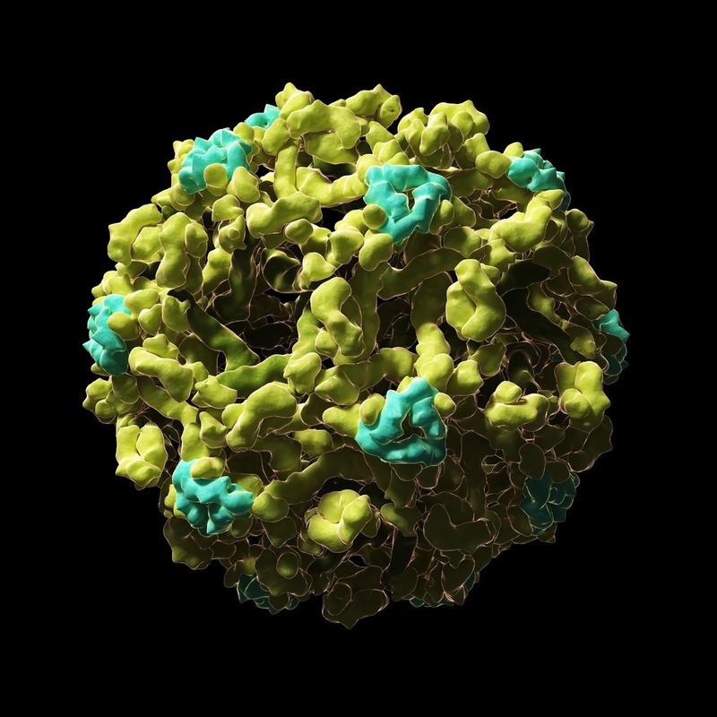
Super-resolution microscopy also plays a part. It breaks the diffraction limit barrier, a longstanding challenge in microscopy. These microscopes can now visualize viral particles at nanometer resolution. They provide insights into sub-viral structures critical for vaccine development.
Adaptive optics is another feature to celebrate. It corrects distortions in images caused by specimen irregularities. This technology ensures that the images of viruses are not only high-resolution but also true to their actual forms.
In 2025, these microscopes are more user-friendly and cost-effective. They make virus visualization more accessible to labs around the world. This democratization of technology is crucial. It ensures that diverse research teams can contribute to global virology knowledge. With such progress, we are better equipped to combat viral threats and protect public health.
The Importance of Observing Viruses
Understanding viruses is key to public health. By observing a virus under a microscope, scientists can identify how it spreads and how it affects cells. This knowledge is vital for creating vaccines and treatments. With clearer images, researchers can decipher the virus’s structure, lifecycle, and weak points more effectively.
Advanced microscopes have boosted our ability to fight diseases. They let us see tiny details of viruses that were once invisible. Now, we can detect new viruses faster and respond to outbreaks more swiftly. We can also track changes in existing viruses. This alerts us to potential risks before they become widespread.
Seeing a virus under a microscope helps educate people too. It shows how real and dangerous viruses can be. This encourages better hygiene and support for vaccination programs. Clear visuals of viruses make the need for prevention and research funding more urgent and apparent.
In summary, viewing viruses at this new microscopic level is changing the game. It’s empowering scientists to protect us from current and emerging viral threats. It also brings valuable insights to the public, making us all part of the solution.
Cutting-edge Imaging Techniques for Virus Visualization
Advanced imaging technologies have pushed the boundaries of how we see viruses under microscopes. These techniques are vital for detailed study and understanding of viral infections. They include several sophisticated methods.
One such method is confocal laser scanning microscopy (CLSM). CLSM allows for high-resolution, 3D images of viruses. It helps scientists view the exact position of a virus in a cell. It’s a key tool for understanding viral entry and replication.
Total Internal Reflection Fluorescence (TIRF) microscopy is also noteworthy. TIRF provides a way to look at the interaction of viruses with cell membranes. This is crucial for studying the initial stages of viral infection.
Another technique is Photoactivated Localization Microscopy (PALM). PALM gives ultra-high-resolution images of viruses. It does this by lighting up individual molecules. This lets us see the arrangement of viral proteins.
Stochastic Optical Reconstruction Microscopy (STORM) uses a similar approach. It achieves superb resolution, beyond the limits of traditional light microscopes. STORM images reveal fine details in the structure of viruses.
These methods are just a few examples of how science has advanced in virus visualization. By using these imaging techniques, researchers can monitor a virus under a microscope with great precision. These breakthroughs help us learn more about viral behaviors and how they can cause disease. The insights gained pave the way for new treatments and preventive measures, safeguarding public health.
The Role of Electron Microscopy in Virology
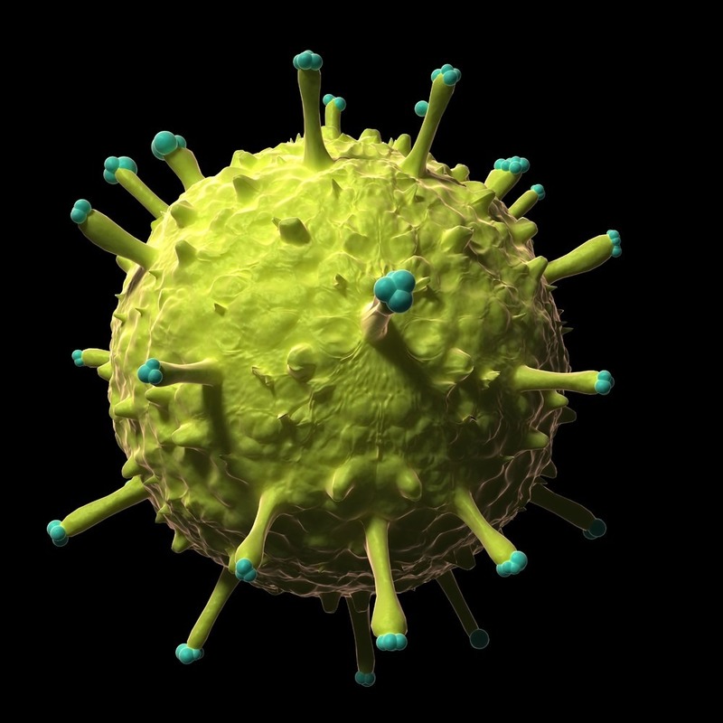
Electron microscopy has reshaped our understanding of viruses. It can visualize viruses at magnifications traditional microscopes can’t match. This is crucial because it reveals the intricate details of a virus under a microscope. Electron microscopes shoot a beam of electrons. These electrons pass through or bounce off the sample, forming an image. With this, scientists can see a virus in high-resolution.
This technology assists in spotting differences in virus structures. This is vital for distinguishing between virus types. Such precise identification helps in creating targeted treatments. Electron microscopy is also effective in visualizing viral assembly. This reveals the stages of how viruses replicate. This knowledge is key in developing antiviral drugs.
Moreover, researchers rely on electron microscopy for observing virus-cell interactions. Understanding how a virus enters and affects a cell guides vaccine development. Visualization tools like electron tomography offer three-dimensional images. These images provide a more complete picture of virus morphology. They are valuable in the fight against viral outbreaks.
In virology, electron microscopy is a powerful tool. It provides images that help decode viral secrets. These insights contribute to improving human health. They are instrumental in our continued battle against infectious diseases.
Innovations in Fluorescence Microscopy
In recent years, fluorescence microscopy has seen significant improvements. These advances have refined how we view a virus under a microscope. Innovations in this field empower researchers to see viral activities with greater contrast and specificity.
One notable innovation is the development of super-resolution fluorescence microscopy. This method surpasses traditional optical limits, enabling scientists to view viral particles with nanometer precision. Detailed imaging of viral components is now possible, providing a deeper understanding of viral mechanisms at the molecular level.
Enhancements in fluorophore technology also play a key role. Brighter and more stable fluorophores have been developed. They improve image quality and allow for longer observation times of viral processes. Researchers can examine the dynamics of virus-cell interaction as they unfold.
In addition, multiplexing techniques have advanced. They allow for simultaneous detection of multiple viral elements. This enables a comprehensive analysis of virus composition and structure.
Another breakthrough comes from live-cell imaging. Fluorescence microscopy can now track the behavior of a virus under microscope in real time. This gives insights into viral replication and spread within living cells, a crucial factor for antiviral research.
These innovations make fluorescence microscopy an essential tool in virology. They expand our ability to study viruses in great detail. As a result, they advance our efforts in developing effective treatments and vaccines. With improved observation, we can better understand and combat viral diseases, safeguarding public health.
Cryo-Electron Microscopy: A Game Changer in Virus Visualization
Cryo-Electron Microscopy (Cryo-EM) has revolutionized the way we see viruses. This advanced technique freezes viruses rapidly. It captures them in their natural state without the need for dyes or fixatives. Scientists call this process ‘vitrification’. Images from Cryo-EM are incredibly clear and detailed. They reveal the architecture of viruses at an atomic level.
Researchers can now ‘slice’ through those frozen samples digitally. By doing this, they obtain 3D images of viruses. These 3D images have changed our understanding of viral structures. For the first time, we see the tiny parts making up viruses. These include proteins that allow viruses to enter host cells.
Cryo-EM stands out in visualizing virus assembly and disassembly. It shows how new viruses form and how they are released from infected cells. This is a crucial step in finding ways to stop viruses from spreading.
The contribution of Cryo-EM to vaccine development is also profound. It helps identify parts of the virus that the immune system can target. This knowledge speeds up the creation of effective vaccines. The technique is already improving vaccine design for diseases like COVID-19.
Overall, Cryo-EM is a powerful tool. It gives us high-resolution images of a virus under a microscope. With Cryo-EM, scientists worldwide are making fast progress. They are understanding and fighting viruses better than ever. Cryo-EM is indeed a game changer in the field of virology.
The Impact of High-Resolution Virus Imaging on Public Health
The advances in virus imaging technology, particularly high-resolution methods, have a profound effect on public health. Observing a virus under a microscope with such precision allows for quicker and more accurate diagnoses. As we pinpoint the various structures of different viruses, we can better understand their modes of infection and replication.
One of the critical benefits is the development of more effective vaccines and antiviral therapies. High-resolution images help researchers identify key viral proteins that can trigger immune responses. This precision leads to vaccines that are more robust and targeted.
Rapid and precise identification of viruses also enhances our ability to manage outbreaks. With detailed images, health officials can monitor the spread and mutation of viruses more effectively. This is vital for implementing timely and appropriate interventions.
Public awareness and education have also improved due to clearer images of viruses. When the public sees a virus under a microscope in high resolution, the threat becomes more tangible. This can lead to increased acceptance of preventive measures like vaccinations and hygiene practices.
Furthermore, high-resolution imaging is crucial for tracking the evolution of viruses. By observing the minute changes in viruses, scientists can anticipate potential pandemics. They can also develop strategies to prevent or mitigate them.
Overall, the impact of high-resolution virus imaging extends far beyond the research lab. It’s a critical component in safeguarding global health and ensuring we’re prepared for current and future viral threats.
Future Prospects: What’s Next for Virus Imaging Technology?
The future of virus imaging technology holds exciting potential. We expect to see innovations that provide even more detail and insight when viewing a virus under a microscope. Here’s what we might look forward to:
- AI and Machine Learning Integration: Future microscopes might use AI to analyze images faster. They could predict virus behavior, speed up diagnoses, and aid in treatment planning.
- Greater Accessibility and Affordability: Advances may lead to less costly equipment. This would make advanced virus imaging available to more research labs worldwide.
- Enhanced Real-Time Imaging: We anticipate improvements in live-cell imaging. These advances would let scientists watch virus replication as it happens, without any delay.
- Nanotechnology: Nanotech could help us view viruses at an even smaller scale. It might unveil new aspects of virus structure and function.
- Cloud Computing: Cloud-based data analysis might become standard. It would simplify sharing and comparing virus images across the globe.
- Personalized Medicine: High-resolution virus imaging might one day support tailored therapies. Each patient’s virus could be studied to customize treatment plans.
- More Detailed Structural Analysis: We could achieve clearer views of the intricate designs within viruses. This would improve our understanding of their weaknesses and how to target them.
In short, the journey of virus imaging technology is far from over. Each leap forward could mean better public health outcomes and a stronger defense against viral diseases. Anticipation for what’s next is high, and the potential benefits for humanity are vast.


