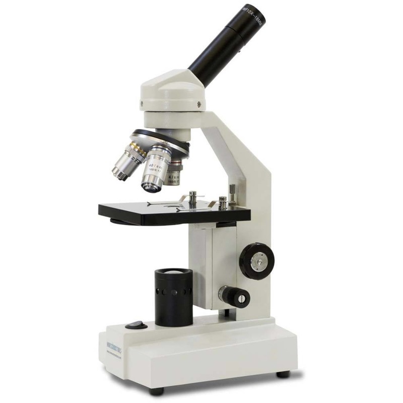
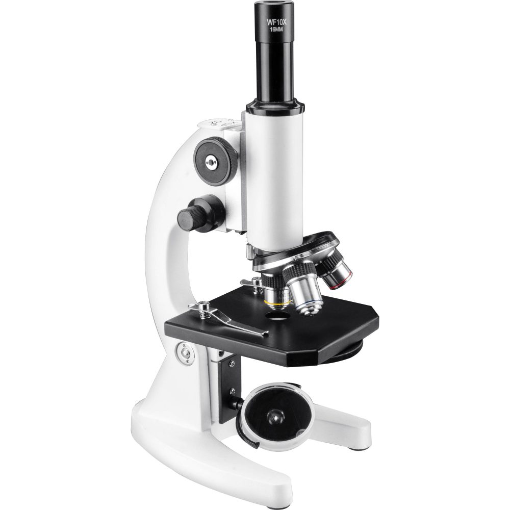
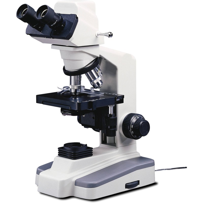
Introduction to Compound Microscopes
A compound microscope is an optical instrument used for viewing objects that are too small to be seen by the naked eye. It’s called ‘compound’ because it combines the powers of multiple lenses to magnify the object. This allows for a closer look at the small details of the specimen being observed.
The compound microscope magnifies the image in two stages. The first magnification occurs through the objective lens, which is near the specimen. The second stage is through the eyepiece or ocular lens, which further magnifies the image to your eye. When you peek through the eyepiece of a compound microscope, you are looking at a greatly enlarged image of the specimen illuminated by light.
This instrument is a staple in scientific labs, used extensively by biologists and researchers. It enhances our understanding of the microscopic world, from examining cell structures to identifying microorganisms. Key to its function are the intricate parts that work together, all of which will be labeled and explained in detail in the coming sections. Understanding these components is crucial for anyone looking to master the use of a compound microscope.
Whether you’re a student, scientist, or curious learner, a deep dive into the anatomy of a compound microscope will equip you with the knowledge to use this powerful tool effectively. In the following segments, we will explore the key components that make up a compound microscope, discuss the optical system including lenses and magnification, delve into the mechanical system focusing on focus and stability, look at the illumination system detailing light sources and filters, outline the steps for sample preparation and stage adjustment, provide tips for maintenance and care, and finally touch upon the technological advancements in compound microscopy.
Key Components of a Compound Microscope
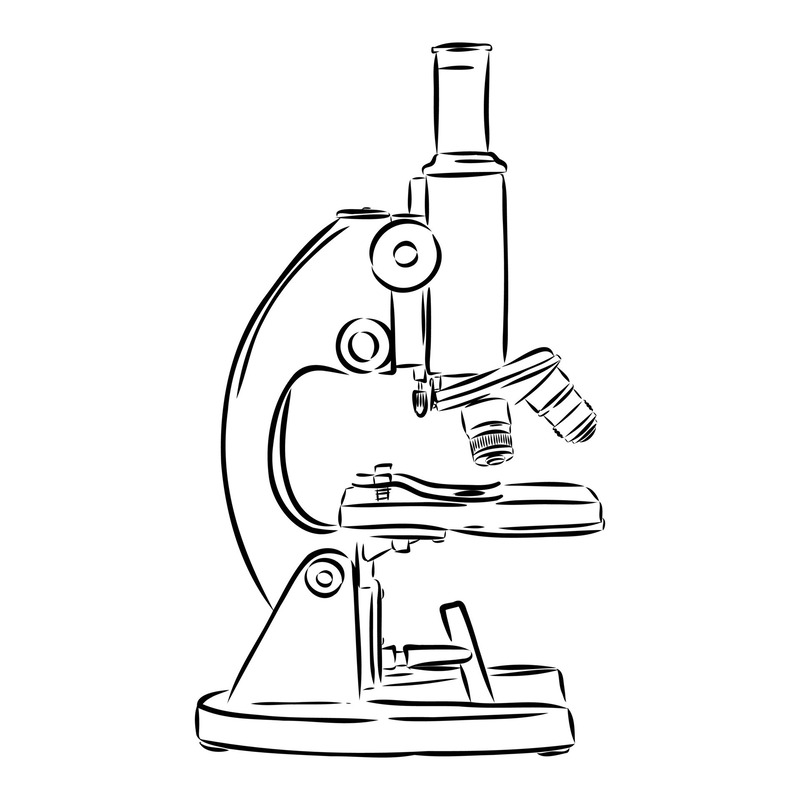
Understanding the key components of a compound microscope is essential for anyone who wants to use this instrument effectively. Each component serves a specific purpose and plays a vital role in magnifying and visualizing specimens. Here, we will break down the compound microscope, labeled to highlight its most critical parts.
- Eyepiece (Ocular Lens): This is where you look through to see the magnified image of the specimen. Typically, the magnification power ranges from 10x to 15x.
- Objective Lenses: Arranged on a rotating turret, these lenses have different magnification powers. Common magnifications are 4x, 10x, 40x, and 100x.
- Body Tube (Head): This connects the eyepiece to the objective lenses and aligns the optical path.
- Arm: The arm supports the body tube and is used to carry the microscope.
- Stage: The flat surface where the specimen slide is placed for observation. Usually, it has clips to hold the slide in place.
- Coarse and Fine Focus Knobs: These knobs adjust the focus of the microscope by moving the stage or the lenses closer or farther from the specimen.
- Illuminator: A steady light source, often LED or halogen, that shines light upwards through the slide.
- Diaphragm or Condenser: These components control the amount of light reaching the specimen. The diaphragm adjusts the light’s intensity, while the condenser focuses the light onto the specimen.
- Base: It supports the microscope and houses the illuminator.
Each component works together to produce a clear, magnified image. When using a compound microscope, labeled diagrams can help users familiarize themselves with these parts and understand their significance. Regular practice combined with this knowledge can lead to precise and insightful observations under the lens.
The Optical System: Lenses and Magnification
The optical system of a compound microscope plays a crucial role in viewing tiny specimens. It uses a combination of lenses to enhance the image that we cannot see with the naked eye. The two main components in this system are the objective lenses and the eyepiece.
Objective Lenses
Objective lenses are the primary tools for magnification in a compound microscope. Labeled with different magnifications, they are found on a rotating turret allowing users to switch between them easily. Typically, these lenses magnify the specimen 4x, 10x, 40x, or up to 100x. When partnered with the eyepiece, they can significantly increase the total magnification.
Eyepiece (Ocular Lens)
The eyepiece, or ocular lens, is what you look through to see the magnified image. It usually adds an additional 10x to 15x magnification to the image projected by the objective lenses. The eyepiece and objective lenses work together to produce a much larger and clearer picture of the specimen.
Total Magnification
To determine the total magnification, you multiply the magnification of the objective lens by the magnification of the eyepiece. For instance, if you are using a 40x objective lens and a 10x eyepiece, the total magnification will be 400x.
Resolution
The resolution, or the ability to distinguish two points as separate, is just as important as magnification. A good optical system doesn’t just enlarge an image, but also keeps it clear and precise. Higher resolution allows for more detailed observations.
Importance of Magnification
Magnification is vital for exploring the details of tiny objects. It allows us to see the intricacies of cells, bacteria, and other microorganisms. By understanding how the optical system works, particularly how compound microscope lenses magnify, you can effectively study the microscopic world.
The Mechanical System: Focus and Stability
Compounding the precision of the optical system, the mechanical system in a compound microscope labeled for focus and stability is crucial for obtaining sharp imagery. Ensuring that specimens are viewed under optimal conditions, this system comprises elements like the coarse and fine focus knobs, the arm, stage, and the base.
Coarse and Fine Focus Knobs
These knobs play a pivotal role in clarifying the specimen image. The coarse focus knob makes rapid adjustments, bringing the object into a rough focus. It’s particularly useful when switching to a higher magnification objective. The fine focus knob, on the other hand, allows for more detailed adjustments, providing a sharper image by moving the stage minutely or adjusting the lenses with precision.
Arm
The arm not only acts as a handle for carrying the compound microscope but also plays a part in maintaining the system’s stability. It firmly connects the body tube to the base, ensuring the alignment of the optical parts remains consistent throughout use.
Stage
As the platform for the slide, the stage’s stability is essential. It generally includes clips or mechanical stage controls that hold the slide securely. By maintaining the slide’s position, it allows for accurate and stable viewing as the focus knobs are adjusted.
Base
Lastly, the base supports the entire structure. Additionally, it securely houses the illuminator in a compound microscope labeled for stability. It provides a dependable foundation that aids in keeping the microscope stable while adjustments are made, minimizing vibrations and potential disruptions.
For users, getting familiar with these mechanical aspects is key to effectively operating a compound microscope. Successful magnification depends not only on the lenses but also on the consistent and precise adjustments that the mechanical system facilitates. Managing the fine balance between focus and stability is what enables researchers to capture images with remarkable clarity.
Illumination System: Light Sources and Filters
The illumination system is a critical part of any compound microscope labeled for efficiency. This system enhances visibility and contrast of the specimen. Let’s take a closer look at its main components.
Light Sources
Proper illumination comes from the light source of the microscope. Commonly, LED or halogen bulbs are used. They provide bright and steady light, critical for clear imaging. LEDs are energy-efficient and produce less heat. Halogen bulbs offer intense illumination that can be adjusted. Choose the light based on your observation needs.
Filters
Filters adjust the light quality reaching your specimen. They can increase contrast or highlight specific structures. Different filters serve different purposes. Some are used for reducing glare, others for enhancing colors. Always match the filter with the type of observation you are conducting.
An effectively labeled illumination system means less strain on your eyes. It allows for lengthy periods of observation without discomfort. For example, a microscope labeled with a daylight filter can mimic natural light. This reduces eye fatigue during prolonged use.
In summary, the illumination system’s light sources and filters are key for a well-functioning compound microscope. They ensure that what you observe is not just magnified, but also visible in great detail. Regular checks and maintenance of these parts will keep your microscope in optimal condition.
Sample Preparation and Stage Adjustment
Before beginning microscopic observation, proper sample preparation is essential. Here is the process outlined in simple steps.
- Slide Preparation: Place your thin specimen slice onto the slide.
- Specimen Fixation: Often, a cover slip is put over to protect the sample.
- Stage Clipping: Use stage clips to secure the slide on the microscope stage.
- Positioning: Carefully move the stage so the sample is under the objective lens.
- Focusing: Begin with the coarse focus knob for general focus, then switch to the fine focus for a clear image.
Adjusting the stage is a delicate task. Too much force can damage the slide or the specimen. Use gentle movements to adjust the stage left, right, forward, or backward. This makes sure your specimen is always centered for the best view.
To assure accuracy, microscopes often have a mechanical stage. This provides precise control over slide movements. It’s especially useful at high magnifications when the field of view is limited.
Effective sample preparation and stage adjustment are key to successful compound microscope use. They underpin all following observations and ensure your results are as accurate as possible. Remember to handle samples and use stage controls with care.
Maintenance and Care for Compound Microscopes
Taking care of a compound microscope labeled with many parts is key to its longevity and performance. Proper maintenance ensures that your microscope functions accurately every time you use it. Here are essential maintenance and care tips to keep in mind:
- Regular Cleaning: Gently wipe the lenses with lens paper. Avoid using tissue or cloth that might scratch the surfaces. Clean the body and stage with a soft, lint-free cloth to remove dust.
- Avoid Moisture: Store your microscope in a dry place. Moisture can damage the lenses and mechanical parts.
- Cover When Not In Use: Use a dust cover to protect the microscope. This prevents dust from accumulating on the components.
- Handle With Care: Always hold the microscope by its arm and base. Rough handling can misalign the optics or damage its parts.
- Check the Illuminator: Ensure the light source is functioning properly. Replace bulbs when necessary and clean any filters regularly.
- Secure Storage: Keep the microscope in a safe space where it won’t be knocked over or jostled.
- Regular Inspections: Periodically check all moving parts. Ensure the focus knobs and stage controls are working smoothly. If they’re stiff, consult a professional before applying lubricants.
- Professional Servicing: Have your microscope serviced by a professional every few years to maintain optimal performance.
By following these maintenance guidelines, your compound microscope labeled with functionality will operate effectively for years. Teaching proper care should also be a part of any training for those new to using these intricate instruments.
Advancements in Compound Microscopy Technologies
Technological progress has greatly enhanced the abilities of compound microscopes. Here, we will explore recent advancements that have been game-changers in microscopy.
Digital Integration
Modern microscopes often incorporate digital cameras. These cameras capture detailed images directly from the microscope. Users can then save, share, and analyze these images on computers. This digital integration has made it easier to document and discuss microscopic findings.
Software for Image Analysis
Sophisticated image analysis software empowers researchers to quantify what they see. The software can measure cell sizes, count particles, and analyze structures within specimens. This boosts the accuracy and productivity of microscopic investigations.
Improved Resolution
Advances in lens technology have improved the resolution of microscopes. Now, users can observe finer details than ever before. This clarity is crucial for research in fields such as microbiology and genetics.
Enhanced Illumination
LED lighting has revolutionized microscope illumination. It provides a consistent, cool light source. This protects delicate specimens from heat damage. Furthermore, it is cost-effective and energy-efficient.
Fluorescence Microscopy
The development of fluorescence microscopy has expanded the capabilities of compound microscopes. With this technology, specific components of a specimen can be lit up using a fluorescent dye. This makes certain structures stand out, facilitating detailed study.
User-Friendly Design
Ergonomics have played a role in recent designs. Modern microscopes offer more comfort during extended use. Features such as adjustable eyepieces and ergonomic stages minimize strain on the user.
These advancements have opened up new possibilities for exploration and discovery. With each leap forward, the compound microscope labeled as a simple tool evolves. It becomes an even more powerful instrument for science and education.


