History of the Microscope
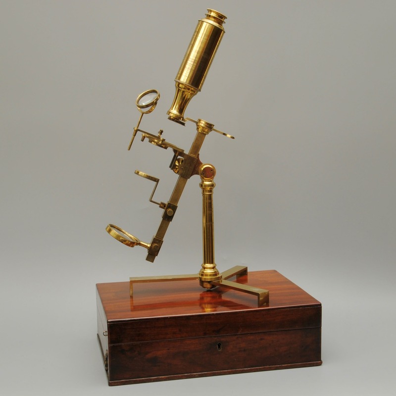
The history of the microscope begins in the late 16th century. Early versions, called ‘flea glasses,’ were simple magnifying devices. However, they lacked the ability to provide detailed views. The first compound microscope, using more than one lens, came to light in the 17th century. This innovation is often credited to two Dutch spectacle-makers, Hans and Zacharias Janssen. They discovered that combining several lenses improved magnification.Learn the microscope definition, its types, how it works, and its applications in science.
The microscope truly evolved after Antony van Leeuwenhoek, a Dutch tradesman. He used self-designed single-lens microscopes to observe the microscopic world. Leeuwenhoek’s microscopes had superb magnification and resolution for their time. His work also paved the way for microbiology. He was the first to see bacteria, free-living and parasitic microscopic protists, sperm cells, blood cells, and much more.
Basic Components of a Microscope
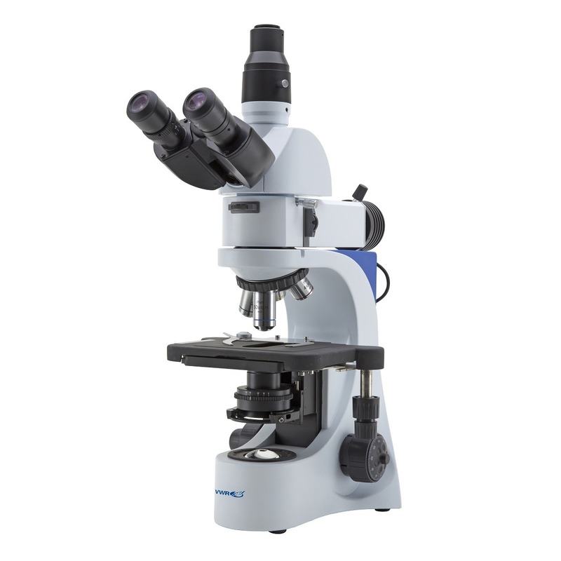
A microscope is a complex instrument. To understand its utility, we must examine its basic parts or components. Each part serves a distinct purpose in magnifying and clarifying the object under study. Here are the fundamental components critical to a microscope’s function:
Eyepiece or Ocular Lens
The eyepiece is where you look through to see the magnified image. It typically possesses a magnification power of 10x or 15x.
Throughout the 18th and 19th centuries, microscope design improved steadily. Innovators such as Robert Hooke and Joseph Lister made significant contributions. Hooke’s book, ‘Micrographia,’ captured the public’s imagination. It included illustrations of specimens seen under the microscope, such as fleas and plant cells. Lister improved the optical quality and reduced the chromatic effect that distorted colors seen through lenses.
In the 20th century, the electron microscope marked a significant breakthrough. The microscope overcame the limitations of light microscopes. It uses electron beams instead of light, pushing magnification and resolution to new levels. Today’s microscopes, as a result of continuous advancements, are powerful tools in science and medicine. They enable us to explore worlds that would otherwise remain invisible to the naked eye.
Understanding the history of the microscope helps appreciate how this essential tool has evolved. It’s interesting to see that from simple beginnings, the microscope has become a cornerstone in scientific discovery. The microscope definition, as we know it today, encompasses a complex and multifaceted instrument. This is thanks to centuries of enhancements and passionate individuals devoted to its development.
Objective Lenses
These are the primary magnifying lenses. Most microscopes have a revolving nosepiece with several objective lenses. Magnifications commonly range from 4x to 100x.
Stage
The flat platform where the slides holding specimens rest. It often has clips to secure the slides in place.
Illuminator or Light Source
Modern microscopes use a built-in light source, usually LED, to illuminate the specimen.
Condenser and Diaphragm or Iris
The condenser focuses light onto the specimen. It’s typically located under the stage. The diaphragm or iris adjusts the amount of light reaching the specimen.
Focus Knobs
These knobs adjust the focus of the microscope. There are typically two: a coarse focus for general adjustments and a finer focus for detailed clarity.
By understanding the microscope’s components, we grasp its capability to bring the unseen into view. The microscope definition is incomplete without acknowledging how these parts work together seamlessly to enhance scientific observation and discovery.Learn the microscope definition, its types, how it works, and its applications in science.
Types of Microscopes
When diving deeper into the microscope definition, it’s crucial to explore the various types available. Different types of microscopes serve unique purposes and are suited for distinct observations.
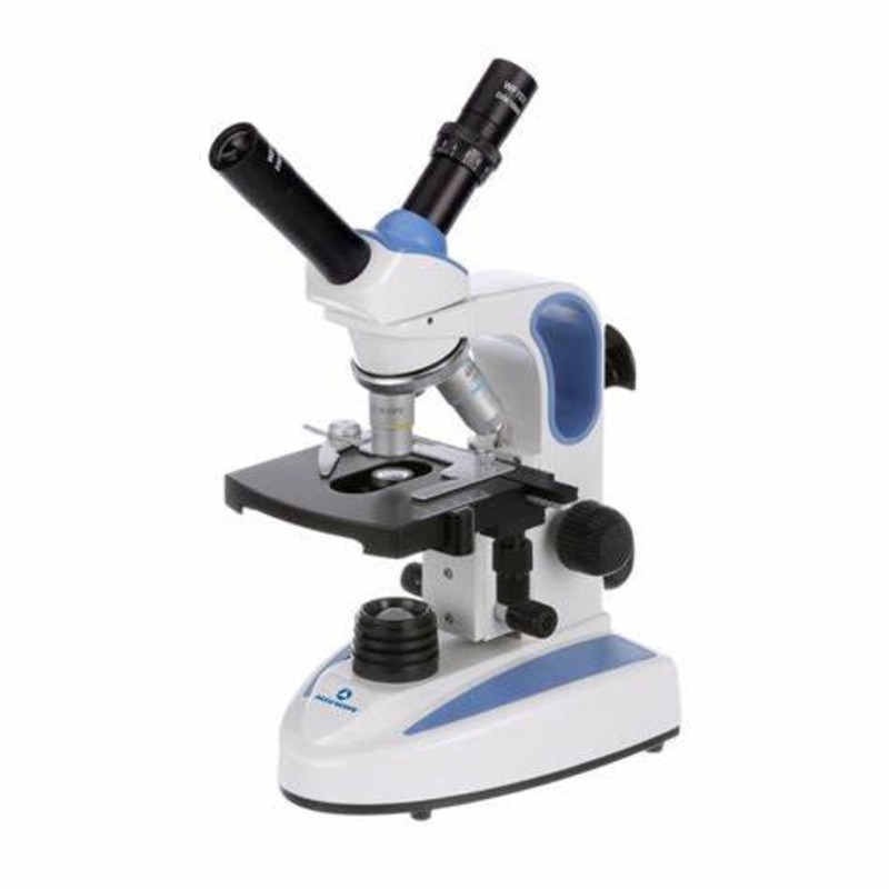
Light Microscopes
The most common type is the light microscope. It uses visible light to illuminate the specimen. It’s perfect for viewing cells, tissues, and other biological samples in high detail.
Electron Microscopes
For ultra-high resolution, scientists turn to electron microscopes. These work with electron beams and are vital for viewing cellular structures or materials at the molecular level.
Scanning Probe Microscopes
At the forefront of nanotechnology are scanning probe microscopes. These devices can image surfaces at the atomic scale, making them essential in material science.
Fluorescence Microscopes
Specialized for viewing samples that emit light when illuminated, fluorescence microscopes are key in molecular biology studies.
Each type of microscope has been tailored to uncover the hidden details of the world around us. They capitalize on different principles, like light, electrons, or scanning probes, to magnify and resolve the minuscule wonders of nature and technology. Selecting the right type hinges on the intended application and the level of detail required.
The Role of Magnification and Resolution
To truly understand a microscope’s purpose, we must grasp two key concepts: magnification and resolution. Magnification refers to how much the microscope enlarges the size of the specimen. Meanwhile, resolution means the microscope’s ability to show detail clearly. In other words, resolution is what allows you to distinguish two points as separate entities. Microscopes with high resolution offer a much clearer and crisper image.
- Magnification is what brings objects closer to our eyes. It helps to see small details that are not visible to the naked eye. But magnification alone is not enough. That’s because you can magnify an image and still see it as blurry.
- Resolution is what gives sharpness to the image. To observe the fine details of a cell or a mineral, you need a microscope with powerful resolution. High resolution will show these details distinctly.
It’s important to note that magnification without adequate resolution would be pointless. The two must work together. For instance, a microscope could magnify a specimen 1000 times. But if its resolution is poor, the image will be of no use. Thus, the perfect balance of both magnification and resolution defines a good microscope.
The microscope definition exemplifies how these instruments are essential for scientific accuracy. Magnification brings us closer to minute worlds. And resolution lays them bare to our scrutiny with clarity. Knowing this helps us appreciate the intricate dance between magnification and resolution in every microscopic investigation.
Microscope Maintenance and Care
Proper maintenance and care are vital for a microscope’s longevity and performance. Here are essential tips to keep your microscope in top condition:
Handle with Care
Always lift the microscope by its base to prevent damage. Avoid touching lenses with your fingers.
Clean Regularly
Use lens paper and special cleaning solutions to gently wipe the lenses. Clean other surfaces with a soft, lint-free cloth.
Store Properly
Cover the microscope with a dust cover when not in use. Store in a dry, temperature-controlled environment.
Check Calibration
Ensure the microscope is correctly calibrated for accurate results. Re-calibrate as needed, especially before significant research tasks.
Inspect Components
Examine all parts periodically for wear and tear. Replace damaged components promptly to avoid affecting the microscope’s function.
Avoid Moisture
Keep the microscope away from liquids. If a spill occurs, immediately dry the affected parts.
Use Correctly
Follow the manufacturer’s instructions for use. This will prevent mishandling and ensure the best possible performance.
By following these maintenance and care steps, your microscope will remain a reliable tool for exploration and discovery. Caring for your microscope means respecting its role in scientific endeavors and the microscope definition of a precision instrument. Regular care ensures your microscope will perform optimally for years to come.
Applications of Microscopes in Various Fields
Microscopes play a crucial role across many fields. From biology to material science, they reveal details invisible to the naked eye. Here are key areas where microscopes are vital:
Biology
In biology, microscopes allow us to study cells, tissues, and microorganisms. They help in understanding diseases and developing new medicines.
Education
Schools and universities use microscopes for teaching. Students learn about cell structures and biological processes.
Medical Diagnosis
Doctors rely on microscopes to diagnose diseases. They examine blood samples, biopsies, and other specimens.
Forensic Science
Forensics experts use microscopes to analyze evidence. This can include fibers, hair, and residue from crime scenes.
Material Science
Microscopes aid in examining materials’ structures. This helps in developing new materials and improving existing ones.
Environmental Science
Scientists study environmental samples with microscopes. They assess pollution levels and monitor ecosystems.
These applications show the versatility of microscopes. They help us explore and learn in diverse scientific domains. The microscope’s definition includes its ability to span different disciplines. This is what makes it an indispensable tool in science and education.
Recent Advances in Microscope Technology
Microscope technology has seen breakthroughs that redefine our microscope definition. Innovations continue to open new frontiers in scientific exploration. Today’s microscopes boast enhancements in magnification, resolution, and user-friendliness. Each advance aims to provide a deeper understanding of the microscopic world. Let’s delve into some of the latest advances in this field.
Digital Imaging and Connectivity
Modern microscopes now often include digital cameras. They capture high-resolution images for analysis. These images can be stored, shared, and reviewed long after initial observation. Some models connect directly to computers or cloud storage, making it easier to collaborate and organize research. Digital connectivity also enables remote learning opportunities, expanding access to this vital tool.
LED Illumination
The shift to LED lighting brings benefits like lower heat output and longer bulb life. LEDs offer consistent and controllable lighting. They reduce the risk of heat damage to samples. This lighting choice is not only more energy-efficient but also provides clearer images. It helps to keep specimens in their natural state during examination.
Enhanced Optical Components
Lenses and mirrors in microscopes are more precise than ever. Advanced coatings and manufacturing processes result in lenses that offer greater clarity. They minimize distortion and provide a true-to-life view of the subject matter. These enhanced optical components also improve resolution, allowing for even finer details to be observed.
Automated Features
Automation in microscopes adds ease of use and repeatability in experiments. Focus, magnification, and lighting can adjust with the touch of a button. Automated systems ensure settings are consistent across multiple uses. This is especially important in research environments where precision is key.
Live Cell Imaging
With new techniques in live cell imaging, researchers can watch cells over time. This captures their natural processes without harm. Time-lapse videos and real-time tracking are now possible. Scientists gain insights into cell life cycles, interactions, and responses to stimuli. These advancements fuel breakthroughs in medical and biological research.
Microscope technology advancements dramatically improve our investigative abilities. They solidify the microscope’s definition as a tool for discovery. With each new development, we step closer to unraveling the mysteries of the micro world.
Choosing the Right Microscope for Your Needs
When you’re in the market for a microscope, picking the right one is vital. Your choice depends on what you need to see and the detail level required. Here’s a guide to help you decide:
- Define Your Purpose: Are you studying bacteria, or inspecting electronic circuits? The object’s size and nature can dictate the type of microscope.
- Consider Magnification and Resolution: If you need to see tiny details, high magnification and resolution are key. But remember, higher isn’t always better. Balance is essential.
- Assess the Light Source: LED lights are common, but for heat-sensitive samples, you might need alternate lighting.
- Digital Capabilities: Do you need to capture and share images? If so, look for a microscope with a digital camera and connectivity options.
- Ease of Use: User-friendly features like automated settings might be important. Ease can speed up your work.
- Budget: Microscopes range from affordable to very expensive. Match your budget with the microscope features you actually need.
- Maintenance and Durability: Consider how often you’ll use the microscope. Frequent use demands a sturdy and easy-to-maintain model.
- Accessories and Upgrades: Check availability for future enhancements. Being able to add components later is useful.
Choosing the right microscope involves understanding your application’s specifics. It’s an investment in your work’s quality. Take time to research and match the microscope’s features to your tasks. This ensures you get a microscope that meets your needs and enhances your scientific endeavors.Learn the microscope definition, its types, how it works, and its applications in science.


