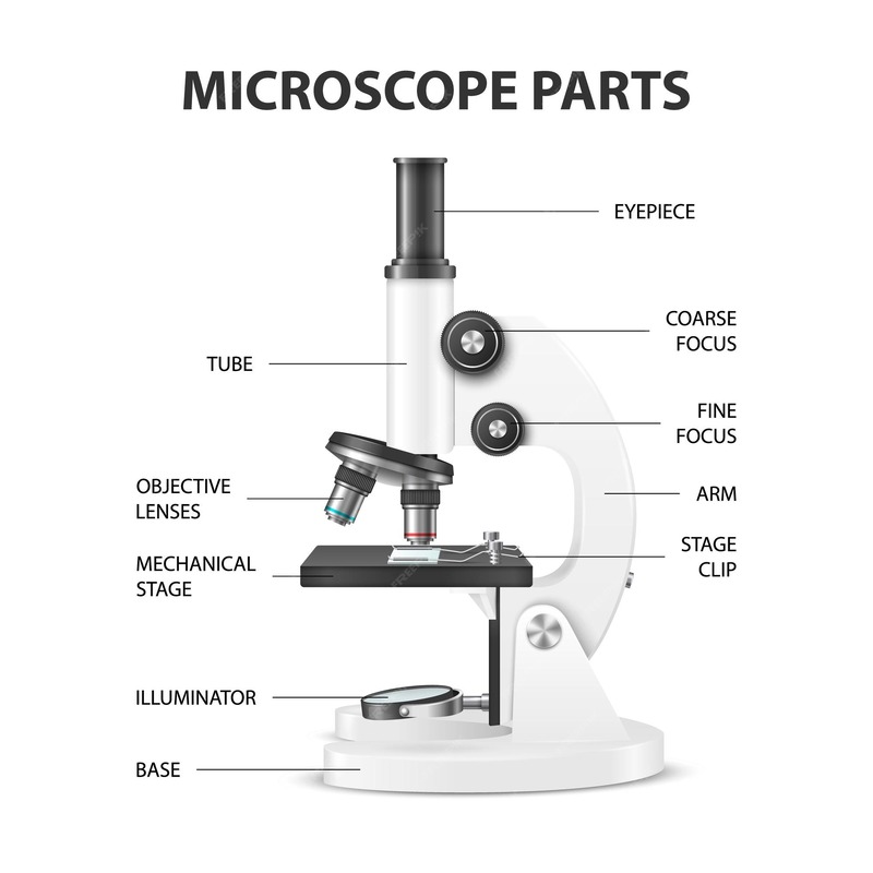
Introduction to Microscope Parts
A microscope comprises various essential parts, each crucial for its functionality. Understanding these components is fundamental for anyone using this instrument, whether in scientific research, education, or any field involving microscopic analysis.Learn about labeled microscope with detailed diagrams and explanation of their parts. Explore how to use a microscope safely and effectively!
Importance of Each Component
Each part of the Labeled microscope serves a specific purpose that contributes to the overall effectiveness of the microscope. Here’s why each component is essential:
- Objective Lenses: These are the primary lenses that magnify the specimen. Without them, the microscope cannot magnify.
- Eyepiece: Also known as the ocular lens, it further magnifies the image formed by the objective lenses and is where you look through to see the specimen.
- Base and Arm: These structural components support the microscope, ensuring stability and durability during use.
- Stage: Holds the specimen in place under the objective lens.
- Illumination system: Provides the necessary light to view the specimen, which is crucial for creating clear images.
- Focusing mechanisms: Include coarse and fine adjustment knobs that help to fine-tune the focus on the specimen, essential for obtaining sharp images.
- Condenser and Diaphragm: These control the light that reaches the specimen, affecting image clarity.
Each Labeled microscope component’s role is interconnected, and understanding these can significantly improve both the usage and maintenance of a microscope.
Objective Lenses
The objective lenses are crucial in a microscope, significantly contributing to its primary function: magnification of specimens. They are positioned close to the specimen and come in various types and magnification powers, each suited for different levels of detail and field depth. Learning about these lenses helps users select the appropriate lens for their particular microscopic needs.
Types of Objective Lenses
Objective lenses range from simple to complex and differ in their capacity to gather light and resolve fine specimen details. Common types include:
- Achromatic Lenses: These lenses minimize color aberration and are great for general lab use.
- Plan Achromatic Lenses: Similar to achromatic lenses but with improved flatness of field.
- Semi-Plan Lenses: Offer better flatness of field compared to standard achromatic lenses but are not as flat as plan achromatic lenses.
- Apochromatic Lenses: These high-end lenses provide the highest resolution and color correction.
Each lens type caters to different requirements, making it essential for microscopic operators to understand their distinctions.
Magnification Powers of Objective Lenses
The magnification power of objective lenses usually ranges from 4x to 100x. Labeled microscope objective lenses are identified by their magnification and numerical aperture, with the following common magnifications:
- Low Power: The 4x objective lens provides a broad view and is ideal for scanning the entire slide.
- Medium Power: The 10x and 40x lenses are used for viewing smaller areas with more detail.
- High Power/Oil Immersion: The 100x lens, used with immersion oil, allows viewing at the highest magnification with intricate details.
Choosing the correct magnification power is critical for achieving the desired level of detail in microscopic examination.
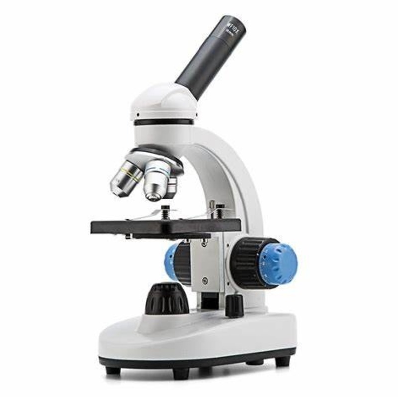
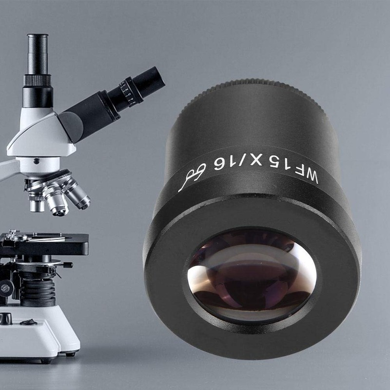
Eyepiece Components
The eyepiece, also known as the ocular lens, Labeled microscope plays a crucial role in magnifying the image of a specimen. It is the lens you look through to observe the magnified image produced by the objective lenses. Proper understanding of the eyepiece is essential for optimizing the use of a microscope.
Role of the Eyepiece in Microscopy
In a Labeled microscope, the eyepiece is the uppermost optical component. It works in tandem with the objective lenses to enhance the magnification of the specimen. The eyepiece typically provides additional magnification of 10x or 15x. This part of the microscope also helps focus the image into the viewer’s eye, ensuring that details are clear and sharp.
Eyepieces can come with or without a pointer. They sometimes include reticles or measurement scales, useful for specific applications like counting cells or measuring microscopic structures.
Magnification and Diopter Adjustment
The magnification Labeled microscope on an eyepiece indicates how much it can enlarge the specimen’s image. Common magnifications range from 5x to 30x but are most often 10x.
Diopter adjustment is a feature found on higher-end binocular microscopes. It allows users to fine-tune the focus specifically for their vision. This adjustment compensates for differences between a user’s eyes, providing a clear image customized to individual sight requirements. Not all microscopes have this adjustment, but it is invaluable for users who need to make precise focus adjustments.
By using the appropriate eyepiece and adjusting the diopter as needed, users can ensure that they obtain high-quality, magnified images that reveal critical details of microscopic specimens.
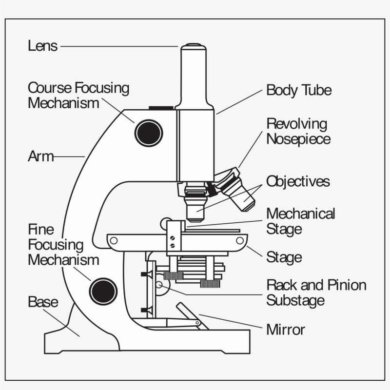
Structural Components
Understanding the structural components of a microscope is as crucial as knowing its optical parts. These components provide the necessary support and stability, ensuring that the microscope operates effectively and safely.
Base, Arm, and Stage
The base, arm, and stage are the primary structural components of a microscope. Each plays a vital role in its functionality:
- Base: The base acts as the foundation of the microscope. It supports the microscope’s weight and houses essential electrical components in many advanced models.
- Arm: The arm connects the base to the head of the microscope. It allows for easy handling and carrying of the microscope.
- Stage: The stage holds the specimen in place during observation. It is typically equipped with clips to secure the slides and is adjustable to facilitate focus.
Adjustments and Supports
Proper adjustment and robust support mechanisms are necessary for obtaining a clear, focused image:
- Microscope Head Adjustment: Allows precise alignment of the optical system with the specimen.
- Mechanical Stage Controls: These are typically knobs that adjust the stage’s position, allowing precise movement of the specimen in both X and Y axes for detailed examination.
These components not only ensure that the microscope is stable and easy to use, but also that it can be adjusted to provide the best possible image of the specimen.
Illumination Components
Understanding the different types of illuminators and their importance is crucial for effective microscopy.
Types of Illuminators Used
Various types of illuminators enhance microscope performance by providing necessary light. Common illuminators include:
- Tungsten-Halogen Lamps: These provide a consistent and powerful light source, ideal for clear observations.
- LEDs: Offer energy efficiency and a cooler temperature, making them suitable for long-term use without overheating samples.
- Fluorescent Lamps: Used for specific applications that require different wavelengths of light.
- Xenon Lamps: Known for their intense and stable illumination, perfect for high-resolution microscopy.
- Mercury Vapor Lamps: Provide bright illumination ideal for fluorescence microscopy.
Choosing the right type of illuminator depends on the specimen and the level of detail required.
Importance of Proper Illumination
Proper illumination is key to obtaining high-quality microscopic images. Here’s why good lighting is essential:
- Clarity and Detail: Adequate light enhances the visibility of specimen details, crucial for accurate analysis.
- Contrast: Proper lighting improves contrast, making it easier to distinguish different structures within the specimen.
- Reduced Eye Strain: Even and adequate lighting reduces eye strain, making microscopy work less taxing.
- Versatility: Different illuminators offer options to work with a variety of samples and conditions.
Ensuring effective illumination enhances the functionality of a microscope and the clarity of images produced.
Focusing Mechanisms
For precise observation, microscopes are equipped with mechanisms that help in focusing on the sample with high accuracy.
Coarse and Fine Adjustment Knobs
To obtain a sharp image, two types of adjustment knobs are utilized:
- Coarse Adjustment Knob: This larger knob makes rapid adjustments, helpful in bringing the specimen into initial focus.
- Fine Adjustment Knob: After coarse focusing, this smaller knob is used for detailed focusing, crucial for clarifying the image.
These knobs work in tandem to attain a crisp view of the specimen under study.
Diaphragm and Condenser Functions
Controlling the light that interacts with the specimen is essential for a clear image:
- Diaphragm: This part regulates the amount of light directed onto the specimen, impacting clarity and contrast.
- Condenser: Position just below the stage, the condenser concentrates light onto the sample, enhancing the resolution and brightness of the observation.
Careful manipulation of the diaphragm and condenser is key to achieving the best visual outcome.
Additional Features
In addition to the main parts, a microscope includes additional features that enhance its functionality and user experience.
Stage Clips and Mechanical Stages
Stage clips are small, yet significant, components of a microscope. They secure the slide in place, preventing movement that can disrupt the view during examination.
Mechanical stages are advanced platforms that allow precise control of the slide position. With knobs to adjust the slide’s X and Y axes, observing specific parts of a sample becomes more straightforward and accurate.
Innovations in Microscope Design
Microscopes are continually evolving, with innovations aimed at improving accuracy, ease of use, and functionality:
- Digital Integration: Modern microscopes often have cameras and software for capturing and analyzing images on computers.
- Environmental Lighting Control: Features like variable LED intensity and color temperature control are now available to cater to different sampling needs.
- Automation: Some microscopes come with automated stage and focus controls, making it possible to conduct scans and analyses with minimal manual input.
These features make modern microscopes more versatile and user-friendly, expanding their applications across various fields of study.


