Introduction
Compound microscopes are vital tools in scientific exploration. They magnify tiny objects using a series of lenses. This instrument has evolved over centuries. Today, microscopes are more advanced than ever. Their purpose remains the same: to reveal the unseen. A compound microscope consists of several essential parts. Each part plays a role in magnification and clarity. Users observe their samples through the eyepiece. They adjust focus and light to get a clear image. Many fields such as biology, medicine, and material science use microscopes. Understanding the parts of a compound microscope is crucial. It helps users to operate it effectively.
In this blog, we will dive deep into the parts of a compound microscope. We will explain how each part contributes to the function of the microscope. From lenses to light sources, every detail matters. Whether you are a student, a scientist, or a curious learner, this guide is for you. Stay tuned as we explore the optical system, mechanical structure, and more.

The Optical System
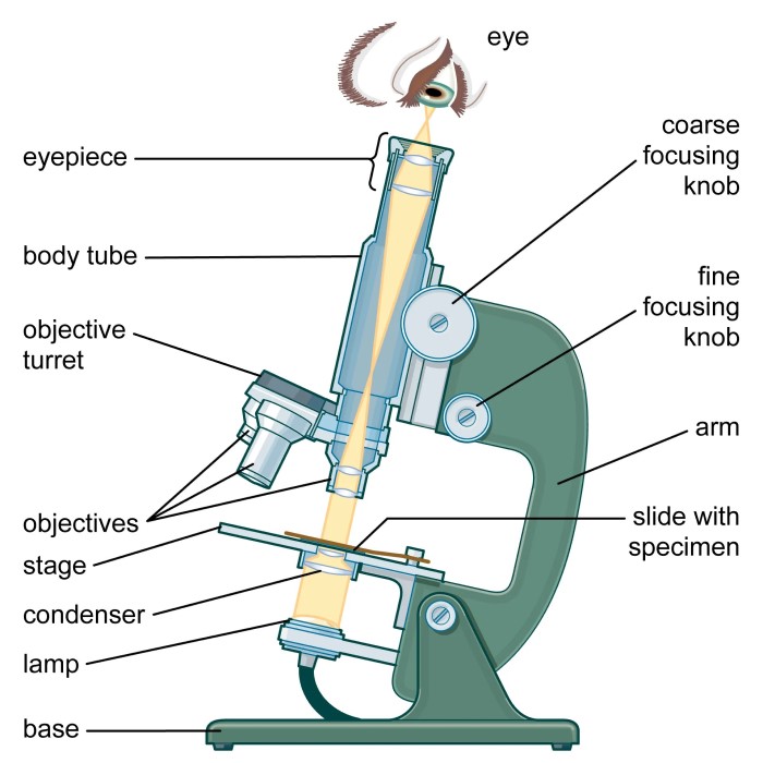
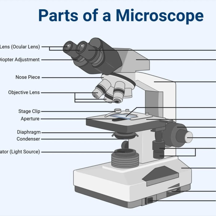
The optical system is a crucial component of the compound microscope. It consists of three main parts: objective lenses, eyepiece or ocular lens, and the tube. Understanding these elements is key to maximizing the instrument’s capabilities.
Objective Lenses
Fundamental Role in Magnification
- Central Component: Objective lenses serve as the essential elements that provide the microscope’s magnification capacity. They are key to transforming a small sample into an observable image that can be analyzed in detail.
Location and Arrangement
- Proximity to Sample: Positioned closest to the specimen, objective lenses directly interact with and gather light emitted or transmitted from the sample being examined. This proximity is crucial for effective light collection and image clarity.
- Multiple Lenses: Typically, a compound microscope features several objective lenses, each designed to offer varying levels of magnification, such as 4x, 10x, 40x, and 100x. This variety allows users to choose the appropriate lens based on the specific requirements of the observation.
Revolving Nosepiece
- Convenient Switching: Most microscopes are equipped with a revolving nosepiece, which allows users to easily rotate between different objective lenses. This feature facilitates rapid adjustments during examination without having to remove the slide.
- Enhanced User Experience: The ability to switch objective lenses seamlessly improves usability and ensures that users can quickly adapt to changing needs during their observations.
Light Gathering and Image Formation
- Light Collection: The primary function of the objective lenses is to gather light from the sample. As light passes through the sample, it carries important information about its structure and composition.
- Magnification Process: Once the light is gathered, the lenses work by bending and focusing it to create a magnified image. This process is crucial for visualizing the intricate details of the specimen, making objective lenses vital for effective microscopy.
Optical Quality and Design
- Lens Construction: Objective lenses are usually made from high-quality optical glass and may have specialized coatings to enhance light transmission and reduce reflections. This quality is essential for achieving clear and precise images.
- Types of Objectives: There are various types of objective lenses, including achromatic, plan, and infinity-corrected lenses, each designed for specific applications in microscopy. Each type offers unique advantages, such as improved color correction or flatness of field.
Impact on Image Clarity
- Resolution Power: The quality and specifications of the objective lenses greatly influence the resolution and clarity of the images obtained. Higher-quality lenses provide finer detail, which is critical for accurate analysis.
- Importance of Cleaning: Proper maintenance, including regular cleaning of objective lenses, ensures optimal performance. Dust or smudges can significantly diminish image quality, making lens care an essential part of microscope operation.
Eyepiece or Ocular Lens
The eyepiece, or ocular lens, is where users actually view the magnified image. It’s located at the top end of the tube. This lens further magnifies the image formed by the objective lenses. Eyepieces commonly have a 10x magnification level. They can be swapped out for lenses with other magnifications, depending on the needs of the user.
Tube
The tube connects the eyepiece to the objective lenses. It’s a key part of the optical system. It holds the lenses in the correct position, maintaining the correct distance between them. This alignment is essential for focusing the image precisely. It lets the user observe the sample in sharp, clean detail. Tubes are typically straight but in some designs, they can be inclined for ergonomic viewing.
The Mechanical System
The mechanical system of a compound microscope is as vital as the optical system. It provides the structural support and precision required for detailed observation.
Stage
The stage is the flat platform where the sample or slide is placed. It must be sturdy and often features a mechanical stage which allows precise movement and positioning of the specimen. This control is crucial for examining different parts of the sample under magnification.
Focus Adjustment Knobs
Focus adjustment knobs are critical parts of a compound microscope for obtaining a sharp image. There are usually two knobs: the coarse and fine focus. The coarse focus knob makes large adjustments, swiftly bringing the sample into the general focus. The fine focus is then used for making smaller, detailed adjustments to the focus, refining the image until it is crystal clear.
Arm and Base
The arm serves as the microscope’s holding area, used for carrying and stabilization. It connects important parts like the stage and optical system. The base provides a firm support for the entire microscope. It helps ensure stability, preventing any movement that could disrupt the view through the eyepiece.
Illumination and Imaging
Proper lighting is essential for microscopy. It enhances clarity and detail in observations. The illumination system in a compound microscope has several fundamental components. These parts ensure that light travels optimally through the sample. This section covers the vital roles of the light source, condenser, and diaphragm or iris.
Light Source
The light source provides the illumination needed to view the specimen. For most compound microscopes, the light is situated below the stage. Common types of light sources include LED, halogen, or fluorescent bulbs. LEDs are popular because they emit less heat and consume less power. Light intensity can affect how clear the image appears. Users can adjust the brightness to suit their viewing needs.
Condenser
The condenser focuses light onto the specimen. It sits just below the stage. A good condenser gathers and directs light rays. This boosts the contrast and resolution of the image. High-quality microscopes often have an adjustable condenser. It allows users to modify light focus for different magnification levels.
Diaphragm or Iris
The diaphragm, or iris controls the amount of light that reaches the sample. It is located within or below the condenser. By adjusting the diaphragm, users can change the light intensity and contrast. This is key for observing finer details in the specimen. A precise diaphragm adjustment is crucial for high-quality imaging. It helps in avoiding either too much or too little light that could obscure details.
Magnification and Resolution
Magnification and resolution are key factors that define a compound microscope’s performance. To discern intricate details and minute structures, one needs a microscope that delivers on both fronts. Let’s delve into how these aspects work and their significance.
Magnification Levels
Magnification levels in a compound microscope relate to how much the image of the specimen enlarges. These levels can vary, typically starting at 40x and going up to 1000x or more. Changing objective lenses alters the magnification. Higher magnifications let users see finer details. However, the clarity of these close-up views depends on the microscope’s resolution.
Resolution Capability
Resolution is the microscope’s ability to distinguish between two closely positioned points. It determines how sharp the image remains at high magnifications. Resolving power is critical; it ensures the magnified image stays clear, not blurry. Microscopes with high resolution capabilities reveal more detail and provide more informative views of specimens. Resolution quality often relies on the quality of the lenses and the wavelength of the light used. Combining high-resolution lenses with proper illumination brings the unseen world into vivid detail.
Sample Preparation and Placement
Proper sample preparation and placement are essential in microscopy. They ensure accurate and clear imaging.
Slide Holder
The slide holder is where you place the glass slide. It keeps the slide stable during observation. Most holders allow for easy slide insertion and removal. The holder’s design ensures the slide remains flat and secure.
Clips or Stage Clips
Clips or stage clips secure the slide on the stage. They prevent movement while you focus and observe. Their purpose is vital for consistent viewing. Clips ensure that the sample doesn’t shift, affecting the image quality.
Maintenance and Care
To keep a compound microscope functioning at its best, proper maintenance and care are essential. Users must follow certain procedures to preserve the integrity and performance of the microscope’s parts. This includes regular cleaning and safe storage.
Cleaning Techniques
Proper cleaning techniques ensure that your microscope stays in top condition. Begin by using a soft, lint-free cloth to gently wipe the body of the microscope. Remove oil and dirt from the objective lenses with a special lens cleaning solution. For the eyepiece or ocular lens, use a dry, soft brush to remove dust. Never touch the lenses with your fingers. Regular maintenance also includes checking for loose screws and misaligned parts. Correct them as soon as possible to prevent damage.
Proper Storage Practices
Storing your compound microscope correctly is as important as cleaning. Keep the microscope covered with a dust cover when not in use. Ensure the storage area is dry and free from temperature fluctuations. Avoid direct sunlight, as this can damage the microscope’s parts. Also, store the microscope with the lowest magnification objective lens in place. This can help protect the more sensitive, high-magnification lenses. By following these storage practices, you extend the life of your microscope.
Advancements in Microscope Technology
As microscopy evolves, new advancements enhance its capabilities. These are shaping the future of scientific exploration.
Digital Integration
Modern compound microscopes now incorporate digital technology. This integration allows for images to be captured and stored on computers. Users can easily share and analyze images. Some models connect directly to software. This software can measure, annotate, and even count specimens on slides. Digital cameras are now common in microscopes. They enable live video streaming. This function aids in education and remote diagnostics. With USB connections, setting up digital integration has become simple. Many microscopes come with built-in digital interfaces. These make the process user-friendly.
Enhanced Imaging Features
Digital Integration
- Modern Technology: Many microscopes now come equipped with digital integration, combining traditional optical systems with advanced digital technologies. This integration allows for seamless connectivity with computers and smartphones.
- Real-Time Viewing: Users can view samples in real time on larger screens, enhancing the collaboration and discussion among researchers during examinations.
High-Definition Cameras
- Clarity and Detail: The incorporation of high-definition cameras significantly improves the quality of images produced by microscopes. This advancement ensures that even the smallest details are captured with clarity.
- Higher Magnifications: As magnification increases, high-definition cameras maintain image quality, allowing for a more detailed examination of specimens without losing clarity in the visuals.
Improved Lighting Systems
- Optimized Illumination: Enhancements in lighting systems lead to better illumination of the samples. Advanced LED lighting options provide consistent and adjustable light intensity.
- Reduced Glare: New lighting technologies minimize glare, which can obscure fine details. This improvement allows for a more accurate assessment of the specimen’s characteristics.
- Contrast Enhancement: Improved lighting enhances the contrast of specimens, making it easier to differentiate between various structures within the samples, resulting in more meaningful observations.
Fluorescence Capabilities
- Specific Dye Tagging: Some modern microscopes are equipped with fluorescence capabilities, enabling users to view samples that have been tagged with specific fluorescent dyes.
- Visualization of Cellular Processes: This feature is invaluable for studying live cells and tissues, allowing researchers to observe dynamic processes and interactions that are critical for understanding biological functions.
Image Processing Algorithms
- Advanced Analysis: Algorithms for image processing are designed to enhance what users can discern through the microscope. They apply various techniques to improve the visuals seen through the lens.
- Pattern Detection: These algorithms aid in identifying patterns and structures that may be difficult to observe with the naked eye. They can enhance the visibility of specific features, allowing for in-depth analysis.
- Quantitative Measurements: Image processing not only improves visual clarity but also enables quantitative measurements of specific structures, facilitating more detailed data collection and analysis.
Expanding Horizons of Scientific Discovery
- Broader Research Opportunities: Advancements in microscope technology are significantly expanding the horizons of scientific discovery. Enhanced imaging features enable researchers to delve deeper into their studies, exploring areas previously inaccessible.
- Interdisciplinary Applications: The improved capabilities of microscopes open up interdisciplinary research possibilities. Fields such as biology, materials science, and medicine are increasingly benefiting from these advancements.
- Increased Learning and Teaching Tools: These technologies serve as powerful tools in educational settings, enriching the learning experience for students and teachers alike.
Conclusion: The Importance of Microscope Components
In summary, the various parts of a compound microscope each play a significant role in scientific exploration. From the sturdy base to the intricate optical components, every aspect of the microscope is designed to work seamlessly together. As you become more familiar with these components, you’ll appreciate the sophisticated engineering behind this essential tool. Understanding the anatomy and functionality of a compound microscope not only elevates your knowledge but also enhances your ability to explore the microscopic world effectively. By mastering these parts, you prepare yourself for a rewarding journey through the fascinating realm of science.


