Introduction
Human ashes, often called cremated remains, offer a unique perspective into the journey of life and death. Understanding their composition can reveal numerous details that are often overlooked during the cremation process. By examining human ashes under microscope, we can unveil intricate structures and chemical compositions that tell a story about an individual’s life, their health, and the impact of cremation on these remains. This article provides insights into the complexities of human ashes, emphasizing the importance of their microscopic examination and what it entails.
What Are Human Ashes?
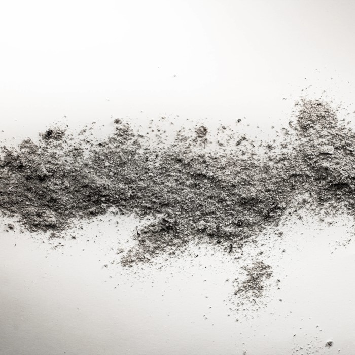
When a body is cremated, what remains is known as human ashes. These are not ashes in the traditional wood-fire sense; instead, they are the powdery residue left after the intense heat has incinerated the body’s organic components. This process reduces the body to bone fragments and a dry powder. The material is often pale gray to white. However, color can vary depending on the temperature achieved during cremation, the composition of the body, and the container used for cremation. Cremated remains are typically ground into a fine dust to ensure a more consistent texture. People often keep these remains in urns or scatter them in meaningful places as a way to memorialize the deceased.
To look at human ashes under a microscope is to peep into a world of unique particles, each with its own story based on the life and body composition of the departed. While from a distance they may seem uniform, up close, the ashes reveal a complex mixture of minerals and compounds. They contain calcium phosphates from bones, but also may include other elements such as sodium, potassium, and magnesium, telling a tale of the life once lived. Examining human ashes under a microscope can help us understand more about the cremation process, the former life of the deceased, and even aid in various types of investigations. The microscopic view of human ashes opens the door to a deeper understanding of the cycle of life and the processes that follow death.
The Process of Cremation
Cremation is a method of body disposal that employs intense heat. The process starts with placing the body in a cremation chamber. Here, temperatures soar to between 1400 to 1800 degrees Fahrenheit. This extreme heat burns away all organic material. Muscles, skin, and organs all vaporize, leaving only bone fragments behind.
During cremation, any metals in the body, such as dental fillings or surgical implants, do not burn. Instead, they are removed from the ash post-cremation using magnets or manual filtering. These metals are often recycled or disposed of according to health regulations.
The remaining bone fragments are not yet the fine powder that many associate with human ashes. To achieve that state, these pieces are placed into a machine that grinds them down. This machine, known as a cremulator, transforms the bone into the uniform, fine dust that families receive.
From start to finish, cremation typically takes two to three hours. However, the entire process, including the cooling and grinding of bones, can take up to seven hours. The final product is roughly four to six pounds of ashes for the average adult. These ashes are the material that, under a microscope, reveal the secrets of our bodily composition and the life once lived.
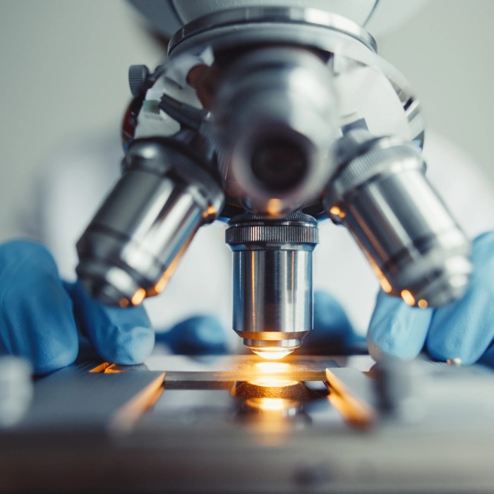
Analyzing Ashes Under the Microscope
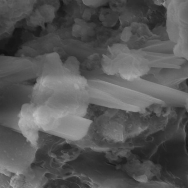
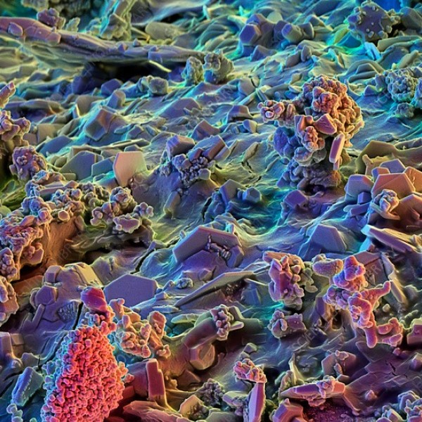
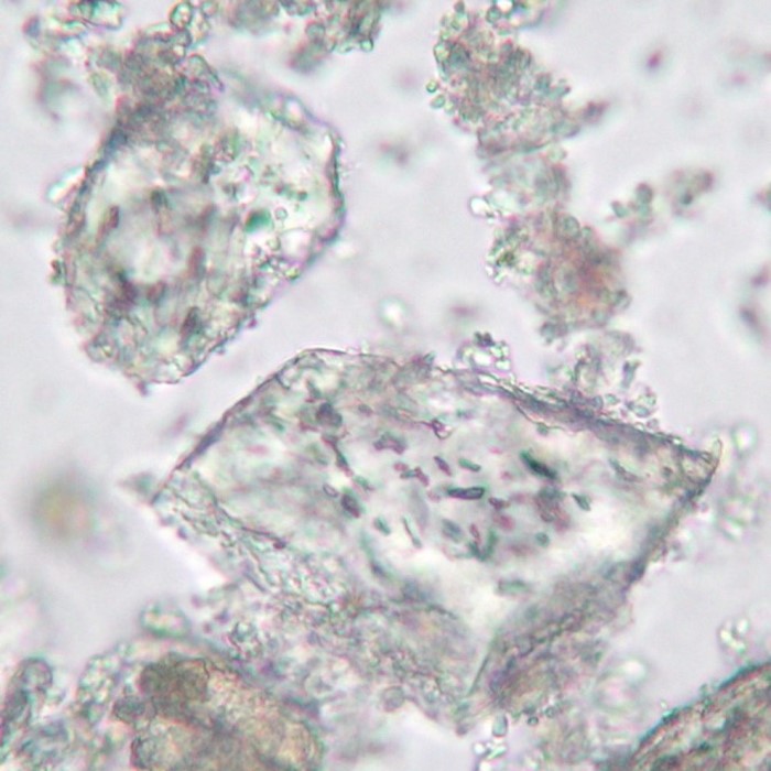
When we examine human ashes under the microscope, an unseen world becomes visible. Microscopy allows us to observe the tiny particles that make up the remains. Here, bone fragments show their unique shapes and sizes, a testament to the high temperatures of cremation. Small, mineral-like structures reflect the once complex biological composition of the human body. Through this lens, we do not just see dust; we see history and biology intertwined.
Under magnification, the fine, uniform appearance of ashes gives way to a diverse landscape. Tiny fractures and crystalline structures within the particles reveal the intense heat’s transformational effects. Variations in colors and textures indicate different temperatures and materials present during cremation. Sometimes, specks of metals from prior medical procedures become visible, having resisted the cremation flames.
Studying human ashes under microscope not just serves as a tool for understanding the physical changes post-cremation. It also helps forensic experts in their investigations, providing clues about a person’s life and the circumstances surrounding their death. As such, microscopic analysis plays a crucial part in modern cremation services and forensic science, allowing for a detailed reading beyond what the eye can perceive.
The Composition of Cremated Remains
When we delve deeper into the composition of cremated remains, we unlock a microscopic atlas of elements. Upon analyzing human ashes under microscope, we find they consist largely of bone-derived minerals, predominantly calcium phosphates. This mineral is what gives bones their hard and durable qualities. But the story doesn’t end there.
Alongside calcium phosphates, one would often find traces of salts such as sodium and potassium. These minerals are residuals from the body’s soft tissues. Magnesium, another element regularly identified in human ashes, plays a role in over 300 biochemical reactions in the human body during life. These remaining salts and minerals form as a result of the high-temperature incineration of organic matter.
Interestingly, the color and texture of human ashes can vary. Variations may tell us about the temperature of the cremation or the presence of unique elements within the body. A lighter hue, for example, could suggest a hotter cremation process. In contrast, darker tones might relate to specific bodily elements that don’t fully break down under extreme heat.
In a closer look, it becomes evident that these remains are not mere dust. They are a testimony to a person’s physical life, bearing the chemical footprints of their existence. The composition of human ashes under a microscope reveals this rich tapestry of minerals. By studying these remains, we gain insight into the life once lived and the enduring materials that last beyond it.
Unique Characteristics in Ash Particles
When we observe human ashes under microscope, we find each particle is distinct. Unique characteristics of ash particles become clear, despite their initial appearance of homogeneity. Under magnification, the individuality of each fragment shines through, shaped by the life of the person and the cremation conditions.
The particles often show pitted surfaces and irregular shapes, results of intense heat. This pitting and shaping are effects of the temperatures within the cremation chamber, leading to an array of particle structures. Moreover, the ashes can reflect the person’s diet, lifestyle, and even the medicines they took. For example, our cells accumulate certain heavy metals over time, and these may be detectable in the ashes.
Microscopic analysis can sometimes identify particles of unique composition. These could stem from prosthetics, dental work, or other surgical implants that were not fully incinerated. The presence of such metals speaks to the medical history of the deceased.
In looking closer, we may also notice subtle color variations. These hues are traces of chemical reactions during the cremation process. Lighter colors tend to indicate a higher cremation temperature, whereas darker shades may be pointers to specific materials affected differently by the heat.
In essence, each set of ashes tells its own story. It’s a modern-day puzzle where we piece together fragments to understand a past life. The uniqueness of ash particles contributes to the rich narrative that is left after a person passes, offering a final glimpse into their journey through life.
Technological Advancements in Microscopy
Exploring human ashes under microscope has become more insightful due to technological progress in the field of microscopy. Advanced tools have enhanced the level of detail and accuracy in studies involving cremated remains. High-resolution imaging provides clearer views of microscopic structures that were once too small to see. This leap in clarity allows us to see the intricate patterns and textures of each ash particle.
Modern microscopes now come with digital interfaces. These interfaces make it easier to capture and share images. With such technology, researchers can analyze ashes remotely and collaborate on findings. This has broadened the scope of examination and understanding of human ashes.
One of the most significant advancements is the use of electron microscopes. Compared to traditional light microscopes, electron microscopes have a much greater magnification power. They can reveal ultra-fine details of the mineral content and structure of ash particles. These details might include tiny fractures and crystalline formations that tell more about the high temperatures during cremation.
Another notable development is the use of spectroscopy aligned with microscopy. This technique allows scientists to identify the elemental composition of the ashes. The ability to detect and measure elements like calcium, sodium, and magnesium provides a deeper understanding of the person’s life.
Lastly, imaging software has become more sophisticated. It enables more detailed analysis and 3D reconstruction of particles. This software helps to provide a better understanding of the extraordinary transformation human remains undergo in cremation.
These technological advancements in microscopy have deepened our appreciation for the intricate details seen in human ashes under microscope. With these tools, we continue to expand our knowledge on the remnants of life’s past.
The Importance of Ash Analysis
Ash analysis plays a crucial role in multiple disciplines, carving its niche as an invaluable practice in modern science and cultural respects. Here are the key reasons why analyzing human ashes under a microscope is significant:
- Identity Verification: In forensic science, ash analysis assists in confirming the individual’s identity. Minute details observed can help match remains to missing persons or verify declared identities.
- Historical Insights: The intricate details in ashes reveal historical data, providing clues about a person’s lifestyle, diet, or environmental impacts on their life.
- Health and Medical Research: By studying the elemental composition of ashes, researchers can learn about the prevalence of certain diseases or the long-term effects of medications and prosthetics.
- Cremation Process Improvement: Understanding how different temperatures and conditions affect the remains aids in refining the cremation process for more uniform and dignified results.
- Environmental Impact Assessment: Analyzing ashes can show the presence of heavy metals or other harmful substances, thus allowing for better environmental regulations in cremation practices.
The significance of human ashes under microscope examinations extends beyond scientific inquiry. It encapsulates emotional and spiritual dimensions for those seeking connection with the deceased, offering closure and a tangible link to loved ones. Ultimately, ash analysis reinforces the respect and honor due to individuals even after they have passed, ensuring their stories and legacies are acknowledged and preserved.
Ethical and Legal Considerations
Analyzing human ashes under a microscope not only unlocks mysteries of our physical being. It also raises ethical and legal issues. It is important to handle such research with sensitivity and respect for the deceased and their families. Any analysis must comply with laws and regulations. Privacy concerns are especially key. Researchers must protect the identity of the individuals involved.
Legal guidelines govern the handling and examination of human remains. These rules vary by region and must be followed. Ethics demand transparent communication with the deceased’s family. This ensures they understand the purpose and outcome of any examination. Any findings must be handled with the utmost care. This avoids misuse of sensitive information.
In research, informed consent is critical for using human ashes. When possible, the consent of the next of kin is sought. This respects the individual’s dignity after death. The study of ashes often aids in forensic investigations. Here, legal standards are strict. They ensure evidence is collected and used lawfully.
Ethical considerations also extend to the handling of metals and other materials found in the ashes. These materials, once personal, are treated with respect. They are often recycled or disposed of safely according to health guidelines.
In conclusion, studying human ashes under microscope reveals much about our biological history. But researchers must navigate ethical and legal terrain with care. Respecting the deceased while advancing science is a delicate balance. With proper care, we can continue to learn while honoring the memory of those who have passed.
Conclusion
Exploring human ashes under the microscope leads to a richer understanding of what remains after death. It is not merely a scientific exercise; it is a profound journey that combines insights about health, personal legacy, and environmental implications. By delving into the analysis of ashes, we unveil the intricate details that make up the story of any individual’s life.
The examination of cremated remains has the potential to inform medical research, facilitate environmental discussions, and foster connections for grieving families. As we continue this journey of discovery, we not only honor those who have passed but also enhance our understanding of life itself.
In summary, studying human ashes under a microscope reveals intricate details about human anatomy and personal histories, offering paths for healing, understanding, and reflection on our shared humanity and the legacies we leave behind.


