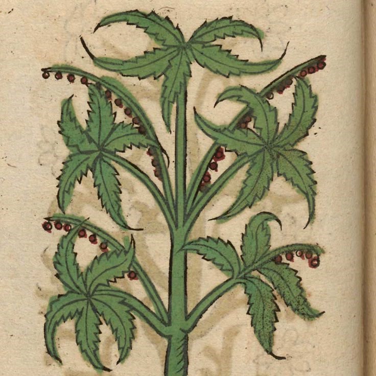
The Cannabis Plant: An Overview
The cannabis plant has long intrigued scientists and enthusiasts alike. A closer look, specifically with the aid of a microscope, reveals a complex structure teeming with life and biochemical activity. When discussing the cannabis under microscope, we’re not just talking about the plant in its entirety. Instead, we explore the intricate details of its anatomy that define its potency, aroma, and therapeutic value.
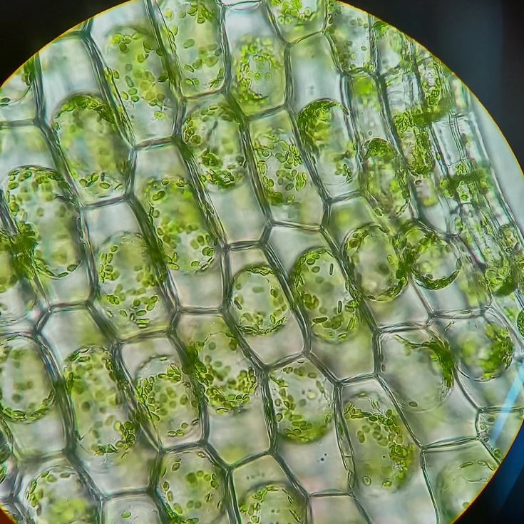
At a macroscopic level, cannabis presents itself with the usual parts: roots, stems, leaves, and flowers. However, as we zoom in, a new world unveils itself. Under the microscope, the plant’s surface is home to trichomes, tiny outgrowths where essential compounds such as cannabinoids and terpenes are concentrated. These trichomes resemble miniature crystalline mushrooms and are the epicenter of the plant’s power.
To fully appreciate what makes cannabis so unique, one must understand its anatomy under magnification. By studying cannabis under microscope, we gain insights into the plant’s properties that are not perceptible to the naked eye. The microscopic examination of the cannabis plant is not just for the scientifically curious. It is also crucial for breeders, researchers, and medical professionals who seek to understand and leverage its potential.
Next, let’s delve into the microscopic anatomy of cannabis to discover what lies beneath the surface, and why these tiny components are pivotal to the plant’s function and our use of it.
Zooming In: The Microscopic Anatomy of Cannabis
When we examine cannabis under microscope, a miniature universe emerges. This detailed view reveals the plant’s essence and its complex internal structures. Let’s take a closer look at the microscopic anatomy of cannabis and understand the critical elements that make this plant unique.
At the heart of cannabis’s microscopic world are its cells and tissues, which form the building blocks of the plant. Through a microscope, the cell walls, nucleus, chloroplast, and other cellular components become visible. These parts are essential for the plant’s growth and the synthesis of its chemical compounds.
One cannot overlook the trichomes, which are tiny, crystal-like outgrowths on the surface of the leaves and buds. Cannabis under microscope shows that trichomes are more than just decorative. They house the plant’s cannabinoids and terpenes, the substances that give the plant its medicinal and recreational properties.
The vascular system of the plant, consisting of xylem and phloem, is also seen under magnification. This system distributes water, nutrients, and the chemical compounds throughout the plant. Stomata, the small openings on the surface of the leaves, are visible too. They play a vital role in gas exchange, allowing the plant to breathe.
A microscopy journey through a cannabis plant also includes discovering the complex structures of the roots, which absorb water and nutrients from the soil. Beneath the soil surface, the roots form a network critical for the plants’ survival and health.
Understanding the microscopic anatomy of cannabis opens up a world of insights into how this plant functions and develops its potent properties. This knowledge is valuable for botanists, cultivators, and consumers alike, looking to grasp the full potential of cannabis.
Trichomes: The Powerhouses of Potency
When observing cannabis under microscope, trichomes demand special attention. These tiny structures hold the key to the plant’s strength. Trichomes appear as shiny, sticky crystals that cover the buds and leaves. They serve as mini factories, making cannabinoids and terpenes. These compounds create the effects and scents we associate with cannabis.

Trichomes come in different types: bulbous, capitate-sessile, and capitate-stalked. Each type has a unique role and appearance. Bulbous trichomes are the smallest and cover the whole plant. Capitate-sessile trichomes are larger and more common. The biggest are capitate-stalked trichomes. They are easy to see and contain the most cannabinoids and terpenes.
By examining cannabis under a microscope, the trichomes’ potency becomes clear. They protect the plant from harmful UV rays and pests. They also lure pollinators with their aromatic compounds.
Cultivators cherish trichomes for their role in determining potency. As cannabis flowers mature, trichomes change color. This change signals the best time to harvest for peak cannabinoid levels.
Microscopy has made studying these powerhouses possible. It allows us to explore how trichomes function and how we can enhance the benefits of cannabis. For anyone interested in the true power of this plant, trichomes are the focal point.
Cannabinoids and Terpenes: A Closer Look
When we explore cannabis under microscope, cannabinoids and terpenes stand out. These are the compounds most responsible for the plant’s effects. Cannabinoids such as THC and CBD are well-known to many. They bind to our body’s endocannabinoid system, causing various effects. Terpenes, on the other hand, are aromatic compounds. They give cannabis its unique scent profile, and also affect its effects.
Cannabinoids are the plant’s chemical messengers. Under a microscope, they are present in the trichomes. This makes the trichomes look like tiny, sparkling jewels. As the plant matures, the trichomes change. This change can tell us about the quality and potency of the product.
Terpenes serve multiple purposes for the plant. They can repel predators and attract pollinators. When we examine cannabis at the microscopic level, we see these terpenes in the trichomes. Each strain of cannabis has a unique terpene profile. This results in a wide range of aromas and effects.
By understanding the role of cannabinoids and terpenes, breeders can develop new strains. They target specific effects or flavors. Insights from microscopic examination guide the breeding process. This leads to more predictable and effective cannabis products for users.
The study of cannabinoids and terpenes is crucial for medical research as well. It helps in creating targeted therapies for various conditions. Under the microscope, the complexities of cannabis reveal themselves. This allows for a deeper understanding of how this plant can aid in health and wellness.
Cannabis Under the Microscope: Techniques and Technologies
The study of cannabis under microscope uses various techniques and technologies. Experts employ light microscopy and electron microscopy to uncover the plant’s secrets. Light microscopy allows for the examination of trichomes and other surface structures in detail. It uses visible light and lenses to magnify the image of the cannabis plant. But for an even closer look, electron microscopy comes into play. This method uses a beam of electrons, which provides much higher resolution. It reveals the cell’s ultrastructure and the molecular composition of cannabinoids and terpenes.
Advancements in imaging technologies have made these microscopic examinations possible. High-resolution cameras capture the intricate details of the cannabis structure. They show changes in trichome density and composition during growth cycles. Software aids in analyzing images to understand the cannabis phenotype. It also helps in identifying the optimal harvest time based on trichome maturity. These technologies contribute to both research advancements and quality control.

Spectroscopy is another vital technology that complements microscopy. It allows scientists to identify chemical compositions without destroying the sample. This non-invasive technique can determine the levels of cannabinoids and terpenes present. Thus, it is essential for product development and ensuring consistency.
For anyone involved in cannabis cultivation or research, mastering these microscopic techniques is key. They reveal the fine details that make each strain unique. This knowledge helps in improving cultivation methods, creating new strains, and ensuring product safety and quality.
The Science of Strain Differences at the Microscopic Level
Differences in cannabis strains are not just about flavor and potency. They’re also seen microscopically. Each strain has unique cellular structures. These impact the plant’s development and effects. By examining cannabis under microscope, we unveil the variance in trichome density and composition. This gives us a preview of a strain’s properties.
Trichomes are key indicators of strain differences at a microscopic level. They vary not only in size and shape across strains but also in their chemical output. More trichomes often mean more cannabinoids and terpenes. This equates to stronger effects. Lesser trichomes might point to a milder experience.
Furthermore, the arrangement of these microscopic structures can influence a strain’s resilience. Some strains show tighter trichome clusters. These can give better defense against pests. Others may have a sparse layout, making them more vulnerable.
Research into the plant’s genetic makeup aids in understanding these differences. It allows breeders to develop strains with desired traits. For example, high CBD strains for medical use, or high THC for recreational effects. Both are the result of selective breeding influenced by microscopic insights.
In summary, the microscope is a powerful tool in judging strain variety. It helps identify the exact stage of trichome maturity. This guides cultivators to harvest at the optimal time. Such precision works wonders in delivering consistent cannabis experiences. It’s the blend of science and art that satisfies both connoisseurs and patients alike.
Advancements in Cannabis Microscopy and Research
Cannabis microscopy has seen impressive breakthroughs in recent years. Advanced techniques have enhanced our knowledge of the plant’s microscopic world. As we peer deeper into cannabis under microscope, we uncover the subtleties of its anatomy and chemistry.
Key improvements in microscopy have resulted in sharper, more detailed images. They reveal the plant’s cellular structures with better clarity. New staining methods have also been developed. These make specific components within the cannabis cells stand out. Such techniques are crucial in identifying cellular arrangements and the distribution of cannabinoids and terpenes with precision.
Research utilizing these advanced microscopes has led to better strain identification. It also aids in the tracking of growth patterns and health of cannabis plants. Experts now can observe the living plant at a cellular level in real-time. This dynamic perspective offers fresh insights into how the plant reacts to its environment. Consequently, studies have uncovered how factors like light, water, and nutrients affect potency and yield.
Cutting-edge microscopes now come with software that automates measurement and analysis. This leap in technology means faster collection and processing of data. It brings efficiency to research, enabling reproducibility and precise results.
These advancements in cannabis microscopy are not just pushing the boundaries of research. They are reshaping cultivation practices and improving quality control. As we continue to dive deeper into the microscopic aspects of cannabis, our understanding of its power and potential enlarges. This knowledge is indispensable for maximizing the therapeutic and recreational benefits of cannabis.
The Role of Microscopy in Quality Control and Safety
The use of microscopy in cannabis cultivation has become critical for quality control and safety. This is because examining cannabis under microscope allows for close scrutiny of the plant’s physical condition, ensuring that only top-quality products reach consumers. By looking at cannabis at the microscopic level, producers can detect early signs of mold, pests, and other contaminants that could harm users.
Microscopy also plays a key role in verifying the plant’s potency. By assessing the trichome development and structure, cultivators can ascertain the optimal time for harvesting. This precision ensures the consistency and effectiveness of the final product. The density and appearance of trichomes under a microscope provide an accurate gauge for cannabinoid concentration.

In addition to verifying product quality, microscopy is essential for safety testing. Labs employ it to test for the presence of unwanted substances. This includes heavy metals or residual solvents from the extraction process. Detailed images help identify such impurities that might compromise the safety of cannabis products.
Lastly, regulatory requirements now often include microscopic analysis as part of the cannabis testing procedures. This sets a standard for safety, making sure consumers receive clean and potent products. As scrutiny on cannabis products heightens, producers who embrace microscopy are leading the way in quality assurance.
Conclusion
Exploring cannabis under microscope reveals a fascinating world filled with intricate structures and active compounds. This microscopic examination provides insights into essential elements such as trichomes, cannabinoids, and terpenes, enhancing our understanding of the plant’s properties and benefits.
Whether you are a scientist, a grower, or an enthusiastic consumer, appreciating the beauty of cannabis at a microscopic level enriches your experience. Knowledge about these microscopic wonders not only informs your choices but also deepens your appreciation for this remarkable plant. Embracing this educational journey allows us to appreciate cannabis in all its forms, from cultivation to consumption, and everything in between.


