Introduction: The Fascination of Skin Anatomy
The human body is both intricate and fascinating, and one of its most remarkable features is the skin. Often overlooked, skin serves as our first line of defense against the external environment. But, have you ever wondered what does skin look like under a microscope? Examining skin at a microscopic level reveals stunning details about its structure, layers, and vital functions. In this article, we will embark on an exploration of skin anatomy, providing insight into what you can expect to see when viewing skin under a microscope.
The Role of Skin in Human Biology
- Protective Barrier: Skin is the body’s largest organ, and it plays a crucial role in protecting underlying tissues from injury, pathogens, and dehydration. It acts as a barrier, preventing harmful substances from entering the body and maintaining homeostasis.
- Thermoregulation: The skin also helps regulate body temperature. Through sweating and altering blood flow to the surface, the skin works to dissipate heat, ensuring that the body maintains a stable internal temperature.
- Sensation and Communication: Additionally, skin contains numerous sensory receptors that allow us to perceive touch, temperature, and pain. These receptors send signals to the brain, enabling us to react to our environment.
- Vitamin D Synthesis: One of the lesser-known functions of skin is its ability to synthesize vitamin D when exposed to sunlight. This vitamin is crucial for bone health and overall well-being.
Layers of Skin: An Overview
What does skin look like under a microscope? Understanding what skin looks like under a microscope begins with an overview of its layers. The skin consists of three primary layers: the epidermis, dermis, and subcutaneous tissue (hypodermis).
Epidermis
- Definition as the Outermost Layer: The epidermis is the skin’s outermost layer, acting as the primary barrier between the body and external environmental factors. It plays a critical role in protecting against pathogens, chemicals, and physical abrasions.
- Composition of Keratinocytes: The epidermis is predominantly made up of keratinocytes, which are specialized cells that produce keratin. Keratin is a fibrous protein that imparts strength and waterproofing properties to the skin, helping to prevent water loss and protect against external damage.
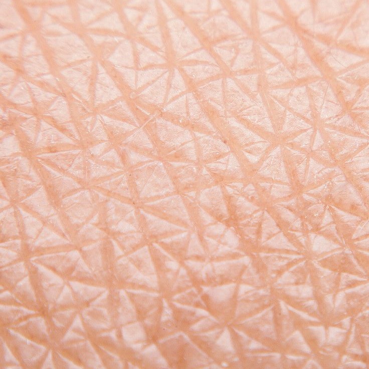
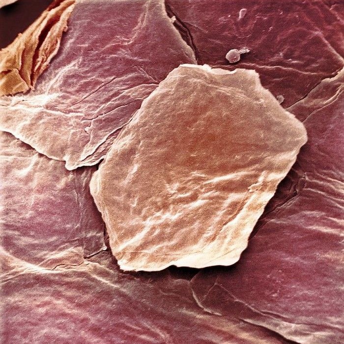
- Layered Structure: Under a microscope, the epidermis displays distinct layers. The outermost layer is the stratum corneum, which consists of dead, flattened keratin-filled cells that gradually shed. Below it lies the stratum granulosum, where keratinocytes further differentiate and begin to lose their nuclei, enhancing the waterproof barrier. Lastly, the stratum basale, the deepest layer, contains actively dividing cells that replenish the outer layers.
- Presence of Melanocytes: Within the epidermis, melanocytes can be observed. These cells produce melanin, the pigment responsible for skin color. Melanin helps protect against the damaging effects of ultraviolet (UV) radiation, and its distribution varies among different individuals, contributing to the diversity of skin tones.
Dermis
- Supportive Function: The dermis is located beneath the epidermis and serves as the supportive layer of the skin. It provides structural integrity and flexibility, which are essential for accommodating movements of the skin.
- Components of the Dermis: The dermis contains various critical structures, including:
- Blood Vessels: These supply the skin with oxygen and nutrients while helping to regulate temperature.
- Lymphatic Vessels: These play a role in immune function and fluid balance by transporting lymph throughout the body.
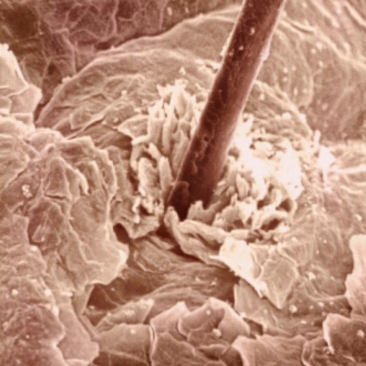
- Hair Follicles: These structures produce hair and are associated with sebaceous glands, which secrete oil to lubricate the hair and skin.
- Sweat Glands: Found within the dermis, these glands help regulate body temperature through perspiration and contribute to the skin’s ability to maintain moisture levels.
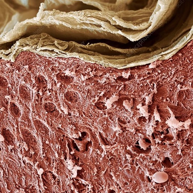
- Microscopic Examination: When viewed under a microscope, the dermal layer reveals dense irregular connective tissue composed of collagen and elastin fibers. Collagen provides tensile strength, while elastin allows for elasticity and flexibility, enabling the skin to return to its original shape after stretching.
Subcutaneous Tissue
- Definition of the Deepest Layer: The subcutaneous tissue, also known as the hypodermis, is the deepest layer of skin. It is composed primarily of fat and loose connective tissue, which provides an essential foundation for the skin above.
- Functions of Subcutaneous Tissue: This layer serves several important functions, including:
- Insulation: The fat cells (adipocytes) within the subcutaneous tissue act as insulation, helping to regulate body temperature and protect internal organs from temperature fluctuations.
- Energy Storage: Subcutaneous fat serves as an energy reserve. The body can draw upon this energy when needed, making it a crucial component of metabolism.
- Cushioning and Protection: The subcutaneous tissue also provides cushioning, acting as a protective barrier for underlying muscles, bones, and organs. This cushioning is vital for preventing injuries from direct impact.
- Microscopic Characteristics: Under microscopic examination, the subcutaneous layer reveals a network of adipocytes that vary in size, depending on the individual’s overall body composition. The loose connective tissue provides structure while allowing flexibility and movement among the layers above.
What Happens When You Look at Skin Under a Microscope?
- Epidermal Details: When looking at the skin under a microscope, the first thing one might notice is the intricate organization of cells in the epidermis. The keratinocytes appear layered, with the outer layers being flattened and more densely packed due to the keratin that has been deposited.
- Diverse Cell Types: The microscopic view also reveals different types of cells present in the epidermis. For instance, melanocytes can be distinguished by their unique shape and pigment production. Langerhans cells, responsible for immune responses, may also be visible, showcasing the skin’s role in immune defense.
- Dermal Complexity: Moving down to the dermis, a microscope reveals a more complex structure. One can observe collagen and elastin fibers that contribute to the skin’s strength and elasticity. Additionally, the presence of blood vessels can be noted, highlighting their role in nutrient delivery and temperature regulation.
- Glands and Follicles: The dermis houses sweat glands and hair follicles, which can be recognized under a microscope. Sweat glands can be pinpointed due to their coils and duct structures, while hair follicles appear as bulbous structures that anchor the hair roots in the skin.
Microscopy Techniques for Skin Analysis
- Light Microscopy: The most common technique used to observe skin anatomy at a microscopic level is light microscopy. It uses light to illuminate the specimen, allowing detailed images of skin layers to be captured. Stains can be applied to enhance contrast, helping to distinguish between various cell types and structures.
- Electron Microscopy: For even greater detail, scientists may utilize electron microscopy, which uses electrons instead of light. This technique provides much higher resolution images, revealing ultrastructural features, such as the arrangement of organelles within skin cells.
- Immunohistochemistry: This technique involves using antibodies to detect specific proteins or antigens in skin samples. It helps researchers identify different cell types and understand their roles in skin health and disease.
The Importance of Understanding Skin Structure
Skin Health
- Understanding Skin Layers: Gaining insights into what skin looks like under a microscope provides valuable information about its various layers, such as the epidermis, dermis, and subcutaneous tissue. Each layer plays a distinct role in maintaining skin health and protecting against environmental aggressors.
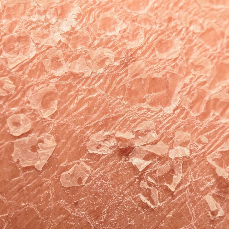
- Diagnosis Improvement: Knowledge of skin anatomy at the microscopic level helps dermatologists and healthcare professionals accurately diagnose skin conditions. For instance, recognizing the characteristic changes in skin layers associated with diseases like psoriasis or eczema can facilitate timely and effective intervention.
- Treatment Plans: With a better understanding of the structural changes that occur in various skin conditions, medical professionals can tailor treatment plans to address these specific issues. This targeted approach can lead to more effective treatments and improved patient outcomes, whether through topical medications, systemic therapies, or lifestyle changes.
- Identifying Skin Cancer: Microscopic examinations are crucial in the early detection of skin cancer. Understanding the typical cellular patterns of healthy and abnormal skin allows for the identification of precancerous lesions and other problematic areas. Early intervention is key in successfully managing skin cancer.
Cosmetics and Dermatology
- Tailored Skincare Solutions: Understanding the microscopic structure of skin enables cosmetic scientists and dermatologists to develop products that cater to various skin types and concerns. Whether addressing oily skin, dry skin, or sensitive skin, this knowledge informs formulations that can penetrate the skin barrier effectively.
- Efficacy of Ingredients: Insights gained from microscopic examinations help in evaluating how different ingredients interact with skin at a molecular level. This understanding is essential for determining which active ingredients can provide therapeutic benefits and enhance skin health.
- Advances in Technology: The cosmetic industry has seen advancements influenced by a deeper understanding of skin structure. Technologies such as nano-encapsulation allow for the delivery of active ingredients into the skin effectively, improving the efficacy of skincare products.
- Personalized Treatments: As research advances, the cosmetic industry is moving toward personalized skincare solutions. By understanding individual skin characteristics, professionals can recommend tailored products that meet specific needs, delivering better results for consumers.
Research and Innovation
- Driving Scientific Discovery: Ongoing research into skin structure at the microscopic level is crucial for uncovering new insights and understanding various skin diseases. These investigations are paving the way for innovations in dermatology and related fields.
- Investigating Skin Cell Behavior: Research focused on skin cell behavior is vital in identifying how different cells interact with one another and respond to environmental factors. This knowledge is instrumental in developing more effective therapies for skin disorders, allowing for more precise medical interventions.
- Regenerative Medicine: Understanding skin at a cellular level is also contributing to advances in regenerative medicine. Research into stem cells and tissue regeneration is illuminating pathways for repairing damaged skin and addressing chronic skin conditions.
- Innovative Therapies: The findings from ongoing microscopic research help pave the way for novel therapies, such as biologic treatments for autoimmune skin diseases and innovative wound healing techniques. Continued exploration in this area holds the potential for breakthroughs in improving skin health and recovery.
Conclusion: A Closer Look at Skin Through a Microscope
In conclusion, exploring what does skin look like under a microscope provides valuable insights into the anatomy and function of this vital organ. By understanding the layers of skin and their cellular organization, we can appreciate skin’s complexity and its critical role in overall health.
With advances in microscopy techniques, researchers can continue to uncover the mysteries of skin. This knowledge not only enhances our comprehension of skin health but also informs everyday skincare choices and treatment options. From protecting our bodies to serving as a diagnostic tool, skin remains one of the most fascinating aspects of human biology. Embracing the scientific exploration of skin opens up new avenues for innovation and understanding in medical science.


