Understanding Viruses and Their Appearance Under a Microscope
What does a virus look like under a microscope? A virus is a microscopic infectious agent that can only replicate inside the cells of living organisms. Unlike bacteria, which are single-celled organisms, viruses are much smaller and more complex in structure. To study them, scientists use powerful microscopes, such as electron microscopes, which allow them to see the tiny details of these pathogens.
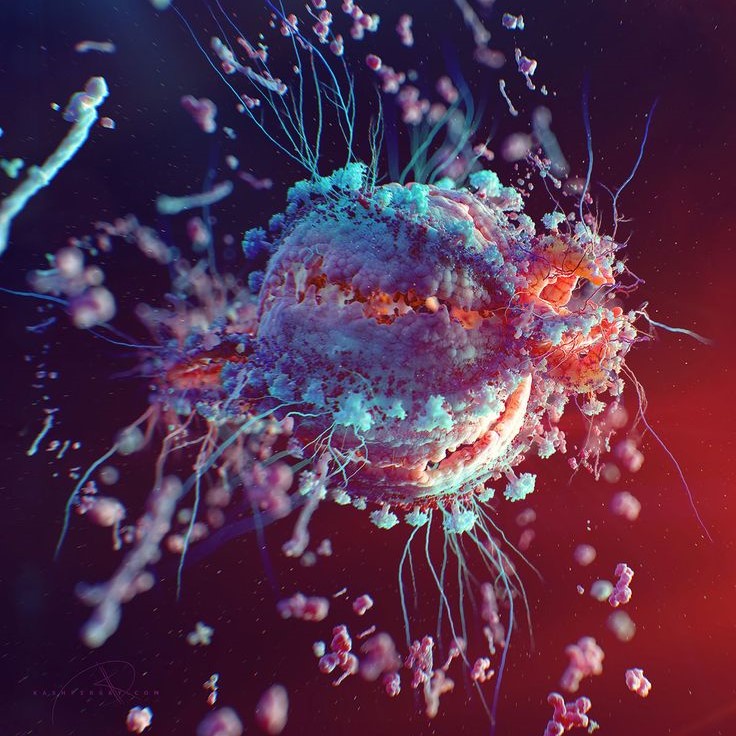
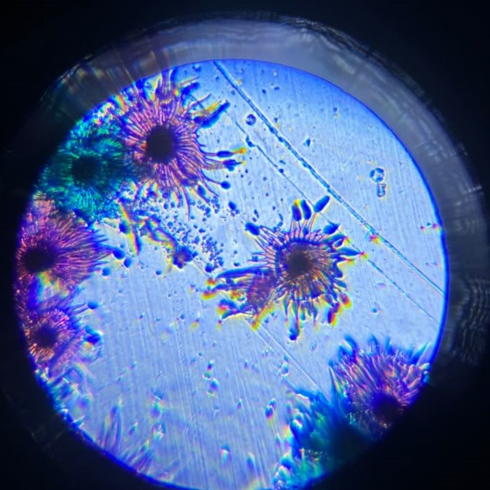
Under a microscope, a virus may appear in various forms depending on its type and the imaging method used. Some viruses have a simple, round shape, while others have more complex structures with spikes or tails. These differences help scientists classify and understand how each virus behaves.

It’s important to note that not all viruses are visible under a regular light microscope. This is because most viruses are too small to be seen clearly with standard magnification. Instead, they require an electron microscope, which uses beams of electrons rather than light to create images.
By understanding what a virus looks like under a microscope, we gain valuable insights into how they infect hosts and how to develop treatments against them.
How Do You Identify a Virus in a Microscope?
Identifying a virus under a microscope involves several steps and techniques. First, scientists must collect a sample containing the virus. This could come from bodily fluids, environmental samples, or infected tissues. Once the sample is prepared, it is placed on a slide and examined under a microscope.
There are two main types of microscopes used for this purpose: light microscopes and electron microscopes. Light microscopes are useful for observing larger particles, but they cannot reveal the fine details of a virus. For this reason, electron microscopes are the preferred tool for studying viruses.
In an electron microscope, the virus appears as a small, often geometric object. Scientists look for specific features such as:
- The shape of the virus (e.g., spherical, rod-shaped, or helical)
- The surface structure (e.g., spikes, capsid, or envelope)
- The size and arrangement of viral particles
These characteristics help determine the type of virus and how it interacts with host cells.
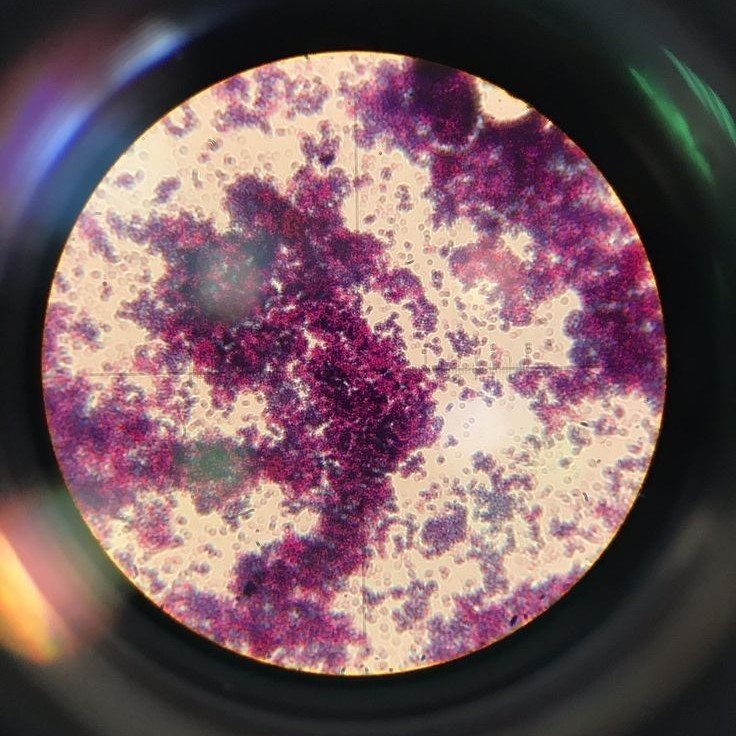
Additionally, staining techniques are often used to enhance visibility. Stains such as iodine, methylene blue, or fluorescent dyes can highlight certain parts of the virus, making it easier to distinguish from other particles.
By using these methods, scientists can accurately identify a virus in a microscope and begin studying its behavior.
What Is the Shape of a Virus Under a Microscope?
What does a virus look like under a microscope? The shape of a virus under a microscope varies greatly depending on the type of virus. Most viruses have one of three basic shapes: spherical (icosahedral), rod-shaped (helical), or complex. Each of these shapes has unique structural features that influence how the virus functions and spreads.
Spherical viruses are the most common. Examples include adenoviruses and polioviruses. Under a microscope, these viruses appear as small, uniform spheres.
Rod-shaped viruses have a long, cylindrical form. They are typically found in plants and some animal viruses. The tobacco mosaic virus is a well-known example of a rod-shaped virus. Under an electron microscope, it looks like a straight, rigid rod.
Complex viruses have irregular or more elaborate structures. These include viruses like bacteriophages, which have a head and tail structure similar to a spaceship. Their complex shape makes them easier to recognize under a microscope.
Understanding the shape of a virus under a microscope helps scientists classify and study these tiny organisms. It also plays a role in developing vaccines and antiviral drugs.
Different Types of Viruses and Their Microscopic Appearance
There are thousands of different types of viruses, each with its own unique appearance under a microscope. Here are some of the most common types and what they look like:
1. Influenza Virus
This virus has a spherical shape with surface proteins called hemagglutinin and neuraminidase. Under an electron microscope, it appears as a small, round particle with spikes on its surface.
2. HIV (Human Immunodeficiency Virus)
HIV has a spherical shape and is surrounded by a lipid envelope. It also has spike-like proteins that help it attach to human cells. These features make it recognizable under a microscope.
3. Ebola Virus
This virus has a long, filamentous shape that resembles a strand of thread. It is often described as having a “snake-like” appearance. Under a microscope, it looks like a twisted, elongated structure.
4. Coronavirus (SARS-CoV-2)
Coronaviruses have a crown-like appearance due to the presence of spike proteins on their surface. These spikes give the virus its name and are easily visible under an electron microscope.
5. Bacteriophages
These viruses that infect bacteria have a complex structure, usually consisting of a head and a tail. Under a microscope, they resemble small, alien-like creatures with a distinct body and tail.
Each of these viruses has a unique shape and structure, which helps scientists identify and study them more effectively.
Electron Microscopy and the Visualization of Viruses
Electron microscopy is the most effective way to see what a virus looks like under a microscope. Unlike traditional light microscopes, which use visible light, electron microscopes use a beam of electrons to produce highly detailed images.
There are two main types of electron microscopes used in virology: transmission electron microscopes (TEM) and scanning electron microscopes (SEM).
Transmission electron microscopes pass electrons through a thin sample, creating a two-dimensional image. This technique is ideal for seeing the internal structure of a virus.
Scanning electron microscopes scan the surface of a sample with a beam of electrons, producing a three-dimensional image. This method is useful for examining the outer shape and texture of a virus.
To prepare a virus sample for electron microscopy, scientists must first fix and stain the specimen. This process helps preserve the structure of the virus and enhances contrast, making it easier to see under the microscope.

Once the sample is ready, it is placed in the electron microscope, and images are captured at high magnification. These images provide a clear view of the virus’s size, shape, and surface features.
Thanks to electron microscopy, we can now see what a virus looks like under a microscope in incredible detail.
Why Is It Important to Know What a Virus Looks Like Under a Microscope?
Understanding what a virus looks like under a microscope is crucial for several reasons. First, it helps scientists identify and classify different types of viruses. This knowledge is essential for diagnosing viral infections and developing targeted treatments.
Second, knowing the structure of a virus allows researchers to study how it interacts with host cells. For example, the shape and surface proteins of a virus determine how it attaches to and enters a cell. This information is vital for creating vaccines and antiviral drugs.
Third, visualizing a virus under a microscope helps track the spread of diseases. By analyzing virus samples from different regions, scientists can monitor mutations and understand how viruses evolve over time.
Finally, learning about the appearance of a virus under a microscope is an important part of education. It helps students and the general public better understand the nature of viruses and how they affect our health.
In short, knowing what a virus looks like under a microscope is not just a scientific curiosity—it’s a key step in protecting public health and advancing medical research.
Common Misconceptions About Viruses Under a Microscope
Despite advances in technology, there are still many misconceptions about what a virus looks like under a microscope. One common misunderstanding is that viruses are always visible with the naked eye or even with a regular microscope. However, this is not true.
Most viruses are far too small to be seen without an electron microscope. Even with a high-powered light microscope, it’s difficult to distinguish a virus from other tiny particles. That’s why scientists rely on electron microscopes to study viruses in detail.
Another misconception is that all viruses look the same. In reality, viruses come in many different shapes and sizes. Some are simple and round, while others are more complex and irregular. This variation makes it possible to differentiate between different types of viruses.
Lastly, there is a belief that all viruses are harmful. While many viruses cause illness, some are harmless or even beneficial.
By correcting these misconceptions, we gain a better understanding of what a virus looks like under a microscope and how it affects our world.
Conclusion
What does a virus look like under a microscope? It depends on the type of virus and the imaging technique used. Most viruses are too small to be seen with a regular microscope, so scientists rely on electron microscopes to observe their structure.
Under a microscope, viruses can appear in various shapes—spherical, rod-shaped, or complex. Their surface features, such as spikes or envelopes, also help identify them. Understanding these details is essential for studying how viruses function and how to protect against them.
We’ve explored how to identify a virus in a microscope, what the shape of a virus looks like, and the importance of electron microscopy in visualizing these tiny organisms. We’ve also addressed common misconceptions and highlighted the significance of this knowledge in science and medicine.
So, next time you ask, “What does a virus look like under a microscope?” remember that the answer lies in the tools we use and the structures we observe. Whether you’re a student, a researcher, or just curious, exploring the microscopic world of viruses is both fascinating and informative.
What does a virus look like under a microscope? It’s a question that continues to inspire discovery and innovation in the field of virology.


