What Does Yeast Look Like Under a Microscope?
Yeast is a single-celled organism that plays a vital role in baking, brewing, and scientific research. When viewed under a microscope, yeast appears as small, round or oval-shaped cells with a distinct structure. These microscopic images help scientists and students understand the biology of yeast, its function, and how it behaves in different environments.
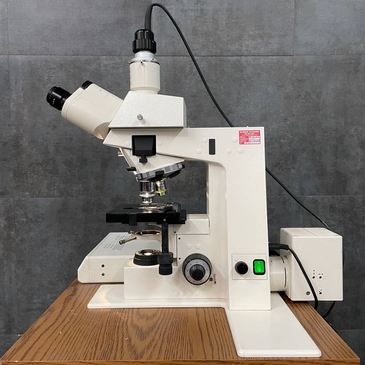
To see what yeast looks like under a microscope, you need to prepare a sample properly. First, mix a small amount of active yeast with water or a nutrient solution. Then, place a drop on a slide and cover it with a coverslip. Finally, use a compound microscope with 40x or 100x magnification to observe the cells.
Under the microscope, yeast cells are usually transparent or slightly opaque. They have a cell wall, cytoplasm, nucleus, and vacuoles. The shape and size can vary depending on the species and growth conditions. Some yeast cells appear round, while others may be elongated or budding.
What does yeast look like under a microscope? It’s important to note that yeast is not visible to the naked eye, but under a microscope, its structure becomes clear and fascinating. This visual guide helps explain what yeast looks like under a microscope and why it’s so important in various fields.
Why Is It Important to See Yeast Under a Microscope?
Seeing yeast under a microscope is essential for understanding its structure, function, and behavior. Scientists, students, and even home bakers benefit from observing yeast at the cellular level. This knowledge helps them monitor fermentation processes, identify contamination, and study microbial growth.
For example, in baking, seeing what yeast looks like under a microscope allows bakers to determine if their yeast is active and healthy. If the cells are swollen, irregular, or dead, the dough may not rise properly. In brewing, observing yeast under a microscope helps brewers ensure the right type of yeast is used and that it’s performing as expected.
In scientific research, studying yeast under a microscope is crucial for genetic studies, microbiology experiments, and drug testing. Yeast is a model organism, and its microscopic features provide valuable insights into cell biology and genetics.
Moreover, seeing what yeast looks like under a microscope is a great way to learn about microorganisms and the invisible world around us. It encourages curiosity and deepens understanding of biological processes.
By using a microscope, you can gain a better appreciation for the tiny yet powerful world of yeast.
What Are the Key Features of Yeast Cells Under a Microscope?
When you look at yeast under a microscope, several key features stand out. One of the most noticeable is the shape and size of the cells. Most yeast cells are oval or spherical, and they range in size from 5 to 10 micrometers in diameter. This makes them much smaller than human cells, which are typically 10 to 100 micrometers in size.
Another feature is the cell wall, which gives the yeast its structure and protects it from environmental stress. The cell wall is rigid and composed of complex sugars like glucan and chitin. This layer helps the yeast maintain its shape and resist damage.
Inside the cell, you’ll see the cytoplasm, which is the gel-like substance where all the metabolic activities take place. The cytoplasm contains organelles such as the nucleus, mitochondria, and vacuoles. The nucleus holds the genetic material, while mitochondria produce energy for the cell.
Additionally, budding is a common feature of yeast cells. Budding occurs when a new cell grows from the side of an existing one. This process is part of asexual reproduction and is a sign of active, healthy yeast.
Lastly, vacuoles are large, fluid-filled structures inside the yeast cell. They help store nutrients and regulate internal pressure, making them important for cell survival and function.
Understanding these features helps explain what yeast looks like under a microscope and how it functions.

How to Prepare a Yeast Sample for Microscopy
Preparing a yeast sample for microscopy is a simple process that requires some basic tools. First, collect a small amount of active yeast—either from a commercial package or a live culture. Mix it with a drop of water or a nutrient broth to create a suspension.
Next, place a small drop of the mixture on a clean glass slide. Make sure the drop is not too thick, as this can make it harder to see individual cells. Use a coverslip to gently press over the sample, spreading the liquid evenly.
If you’re using a compound microscope, start with the low-power objective (10x) to locate the yeast cells. Once you find them, switch to higher magnifications (40x or 100x) for a clearer view. You may also need to stain the sample with methylene blue or iodine to enhance contrast and make the cells more visible.
It’s important to keep the sample fresh and alive during observation. Dead or inactive yeast will not show the same features as living cells. Also, avoid using too much sample, as it can lead to overcrowding and make it difficult to distinguish individual cells.
By following these steps, you can get a clear and detailed view of what yeast looks like under a microscope.
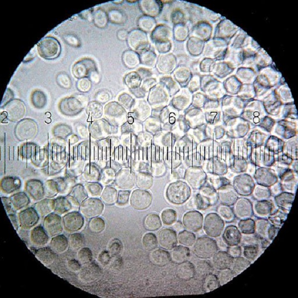
A Detailed Description
Under a microscope, yeast cells appear as small, round, or oval-shaped organisms. They are transparent or slightly translucent, which means they don’t block light completely. This transparency allows researchers to see the internal structures such as the nucleus and vacuoles.
The cell membrane is another visible feature. It acts as a protective barrier that controls what enters and leaves the cell. The cell wall lies just outside the membrane and provides structural support. It is made of polysaccharides and proteins, which give the yeast its rigidity and strength.
One of the most interesting features is the budding process. During this stage, a small protrusion forms on the surface of the cell, eventually growing into a new daughter cell. This is a clear sign of active reproduction and indicates that the yeast is healthy and viable.
You may also notice cell division, where one cell splits into two. This process is essential for yeast growth and is often seen in cultures that are actively fermenting.
In summary, what yeast looks like under a microscope reveals a lot about its structure, activity, and health. Observing these details can help in scientific analysis, quality control, and educational purposes.

Can You See Yeast Under a Microscope Without Staining?
Yes, you can see yeast under a microscope without staining, but staining improves visibility and clarity. Unstained yeast cells are transparent, making it hard to distinguish between the cell membrane, cytoplasm, and other structures. This can limit your ability to study the cell in detail.
However, live yeast cells are easier to spot because they are moving and active. They may appear as tiny, moving dots that bounce around the field of view. This movement is a good indicator that the yeast is alive and working.
Staining techniques like methylene blue or Gram stain help highlight specific parts of the cell, making it easier to see the nucleus, cell wall, and other features. These stains do not harm the yeast significantly, allowing you to study it without destroying the cells.
If you’re using a phase-contrast microscope, you can see yeast cells without any staining. This type of microscope enhances the contrast between different parts of the cell, making it easier to distinguish structures.
In short, while unstained yeast can be seen, staining improves the quality of the image and allows for more detailed observations.
A Visual Comparison
What does yeast look like under a microscope? To better understand what yeast looks like under a microscope, it’s helpful to compare different types of yeast. For example, Saccharomyces cerevisiae, commonly known as baker’s yeast, appears as small, round, and uniform cells. They often budding, which is a sign of active growth.
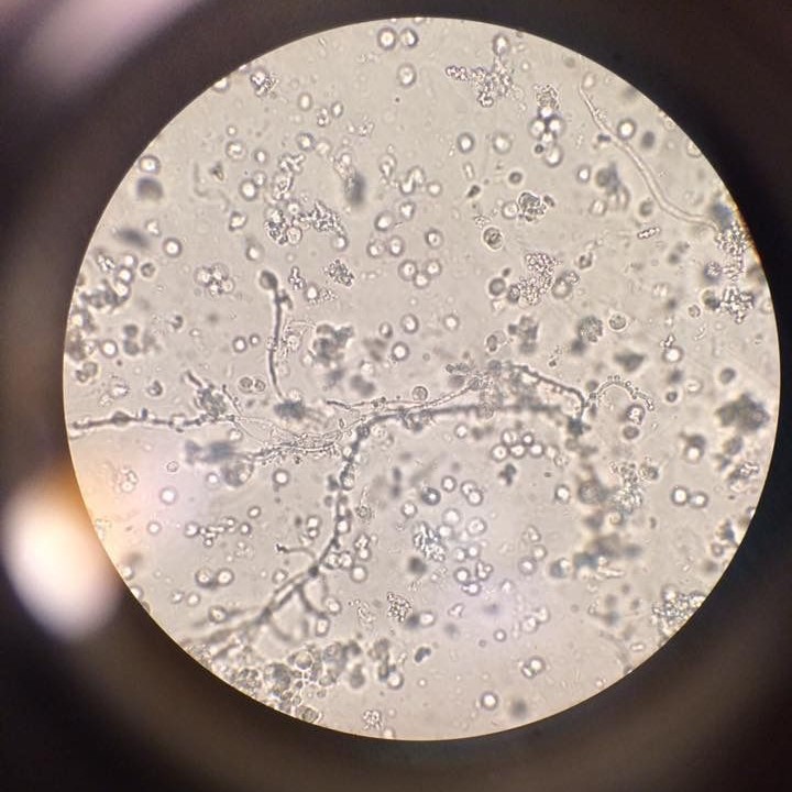
On the other hand, Candida albicans, a type of yeast that can cause infections, has a more irregular shape. It can form filaments or hyphae, which is a key characteristic of this species. These structures are not present in typical baker’s yeast.
Another type, Pichia pastoris, is used in biotechnology. Its cells are slightly larger and more elongated compared to Saccharomyces. This difference in size and shape helps scientists identify and classify yeast species.
Some yeast cultures may also show clumping or clustering, especially when they are in a high concentration. This can make it harder to see individual cells, but it’s a common occurrence in many yeast samples.
By comparing different yeast species, you can better understand what yeast looks like under a microscope and how it varies based on type and condition.
What Are the Differences Between Live and Dead Yeast Under a Microscope?
Live and dead yeast cells show distinct differences when viewed under a microscope. Live yeast cells are active, moving, and have a regular shape. They may be budding or dividing, indicating metabolic activity and growth.
Dead yeast cells, on the other hand, appear deformed, swollen, or fragmented. They may lack a defined shape and do not move. Additionally, they may show signs of ruptured membranes or loss of internal structure.
To check if yeast is alive, you can observe its movement and appearance. Active yeast cells are shiny and well-defined, while dead ones are dull and disorganized. You can also use staining techniques to differentiate between live and dead cells. For instance, trypan blue stains dead cells, making them visible and easy to identify.
In fermentation or baking, knowing whether the yeast is alive is crucial. Dead yeast won’t produce gas, which affects the quality of bread or beer. Therefore, examining yeast under a microscope helps ensure proper usage and performance.
By understanding the differences between live and dead yeast, you can improve your results in both scientific and practical applications.
Final Thoughts on What Yeast Looks Like Under a Microscope
Understanding what yeast looks like under a microscope is a fascinating and educational experience. As we’ve explored, yeast cells are small, round, and transparent, with distinct internal structures that can be observed with the right preparation and equipment.
We’ve also discussed why it’s important to see yeast under a microscope, including applications in baking, brewing, and scientific research. Whether you’re a student, a baker, or a scientist, observing yeast under a microscope offers valuable insights into its structure and behavior.
From budding cells to cell division, each feature tells a story about the life cycle and function of yeast. And by using stains and proper techniques, you can enhance your observations and gain a deeper understanding of this tiny but powerful organism.
Whether you’re looking at live or dead yeast, the visual differences are clear and informative. This guide helps explain what yeast looks like under a microscope and why it matters in science and everyday life.
What does yeast look like under a microscope? It reveals a world of complexity and beauty, showing how **even the smallest organisms play a big role in our daily lives.


