What Is an Electron Microscope and How Does It Work?
How does an electron microscope work? An electron microscope is a powerful tool that allows scientists to see objects at a much smaller scale than a traditional light microscope. Unlike regular microscopes, which use visible light, electron microscopes use a beam of electrons to create highly detailed images of tiny structures. This makes them essential for studying cells, viruses, and even individual atoms.
The basic principle behind how an electron microscope works is simple: it uses electrons instead of light to magnify and observe samples. Electrons have a much shorter wavelength than visible light, which means they can capture more details. As a result, electron microscopes can achieve magnifications up to several million times, far beyond what is possible with optical microscopes.
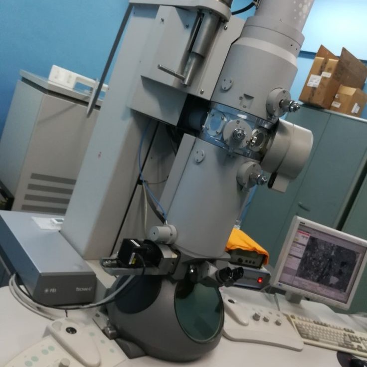
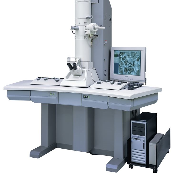
One of the key components of an electron microscope is the electron gun, which produces a stream of electrons. These electrons are then focused using magnetic lenses that act like the glass lenses in a regular microscope. The beam of electrons interacts with the sample, and the resulting signals are detected and converted into images.
Another important part is the vacuum chamber, which keeps the electron beam stable and prevents interference from air molecules. Without this vacuum, the electrons would scatter and lose their energy, making it impossible to get clear images.
In summary, an electron microscope works by using electrons to produce high-resolution images of tiny objects. Its design and technology make it one of the most advanced tools in scientific research.
Why Can Electron Microscopes Only View Dead Cells?
One of the most common questions about electron microscopes is: why can they only view dead cells? The answer lies in the way these microscopes operate. Unlike light microscopes, which can be used on living specimens, electron microscopes require the sample to be dead and fixed before imaging.
This is because the electron beam used in the microscope can damage living cells. The high-energy electrons can cause radiation damage, which kills the cells or alters their structure. Additionally, many electron microscopy techniques require the sample to be dehydrated and coated with a thin layer of metal, such as gold or platinum. These steps are necessary to ensure the sample remains stable under the electron beam and doesn’t degrade during observation.
Furthermore, the vacuum environment inside the microscope is not suitable for living organisms. Most biological samples cannot survive in a vacuum, so they must be prepared in a way that preserves their structure without water or organic material.
In short, electron microscopes are designed for fixed and non-living samples. They provide incredible detail but are not suitable for observing live cells or dynamic processes in real time.
Can an Electron Microscope See an Atom?
How does an electron microscope work? A frequently asked question is: can an electron microscope see an atom? The answer is yes, but with some important conditions. An electron microscope can indeed see individual atoms, especially when using high-resolution transmission electron microscopes (HRTEM). These advanced instruments can resolve features as small as a few angstroms, which is the size of a single atom.
However, there are limitations. For example, electron microscopes require the sample to be extremely thin—often just a few atoms thick. This makes it difficult to study larger or more complex structures. Also, the electron beam itself can damage the sample, especially if it’s delicate or organic.
Moreover, while electron microscopes can detect atomic arrangements, they do not always show individual atoms in a clear, recognizable way. Instead, they often display patterns and structures that represent the arrangement of atoms in a material.
In conclusion, an electron microscope can see an atom, but it requires specific conditions and equipment. It’s a powerful tool for understanding the atomic structure of materials and surfaces.
Why Are Electron Microscopes So Good?
Electron microscopes are considered so good because of their superior resolution and magnification capabilities. Compared to light microscopes, they can see much smaller details, making them ideal for studying nanoscale structures, cells, and even individual atoms.
One of the main reasons they are so effective is their use of electrons instead of light. Electrons have a much shorter wavelength than visible light, allowing for much higher magnification. This makes it possible to see things that are invisible to the human eye.

Additionally, electron microscopes offer excellent depth of field and resolution, which helps in analyzing three-dimensional structures and complex materials. This is particularly useful in fields like materials science, biology, and nanotechnology.
They also allow for chemical analysis through techniques like energy-dispersive X-ray spectroscopy (EDS). This means researchers can not only see the structure of a sample but also determine its chemical composition.
Finally, modern electron microscopes come with advanced software and imaging technologies that enhance image quality and make data interpretation easier. This combination of powerful hardware and smart software makes them indispensable in scientific research.
In short, electron microscopes are so good because they offer unmatched magnification, clarity, and analytical power.
How Does an Electron Microscope Work Step by Step?
Understanding how an electron microscope works involves breaking down the process into several key steps. First, the electron source generates a beam of electrons. This is usually done using a filament or a field emission source, which emits electrons when heated or exposed to a strong electric field.
Next, the electromagnetic lenses focus the electron beam. These lenses are similar to the glass lenses in a light microscope, but they use magnetic fields to control the path of the electrons. This focusing ensures that the beam is directed precisely onto the sample.
Once the beam reaches the sample, it interacts with it in different ways. Some electrons are scattered, while others pass through the sample. These interactions provide information about the structure and composition of the sample.
The next step involves detecting the signals produced by the interaction between the electrons and the sample. Different detectors are used depending on the type of microscope. For example, a screen or camera captures the image in a scanning electron microscope (SEM), while a detector records the transmitted electrons in a transmission electron microscope (TEM).
Finally, the data is processed and displayed as a high-resolution image. This image shows the internal or surface structure of the sample in incredible detail.
By following these steps, an electron microscope provides a clear and detailed view of the microscopic world.
Key Components of an Electron Microscope
An electron microscope consists of several key components that work together to produce high-quality images. One of the most important parts is the electron gun, which generates the electron beam. This beam is made by heating a filament or using a field emitter, both of which release electrons.
Next, the condenser lens focuses the electron beam onto the sample. These lenses are made of magnetic coils that shape the beam and direct it toward the specimen. This focusing is crucial for achieving high magnification and resolution.
The objective lens is responsible for creating a magnified image of the sample. It adjusts the beam to produce a clear and sharp picture, which is then analyzed further.
The sample chamber holds the specimen and ensures it remains stable and undisturbed during the imaging process. It is often evacuated to create a vacuum, which prevents interference from air molecules.
Lastly, the detector system captures the signals produced when the electrons interact with the sample. These signals are then converted into digital images that scientists can analyze.
Together, these components make an electron microscope a powerful and precise instrument for exploring the microscopic world.
Types of Electron Microscopes and Their Functions
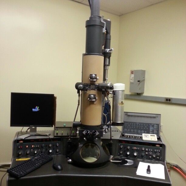
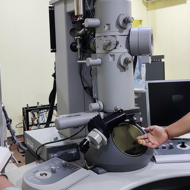
There are two main types of electron microscopes: transmission electron microscopes (TEM) and scanning electron microscopes (SEM). Each has its own unique way of working and is suited for different applications.
A transmission electron microscope (TEM) works by sending a beam of electrons through a very thin sample. The electrons that pass through the sample are then captured and used to create an image of the internal structure. TEMs are ideal for studying cell organelles, crystals, and molecular structures.
On the other hand, a scanning electron microscope (SEM) scans the surface of the sample with a focused electron beam. As the beam moves across the surface, it causes the emission of secondary electrons, which are detected to form a 3D-like image of the surface. SEMs are commonly used for studying textures, surfaces, and topography.
Some advanced models combine both TEM and SEM into a dual-beam system, allowing for both surface and internal imaging. This makes them versatile tools for various scientific studies.
Each type of electron microscope has its own strengths and weaknesses, and the choice depends on the research goal and sample type.
Applications of Electron Microscopes in Science and Industry
Electron microscopes have a wide range of applications in science and industry. In biology, they are used to study cell structures, viruses, and tissues in great detail. Scientists can examine the internal organization of cells and understand how they function at the molecular level.
In materials science, electron microscopes help researchers analyze the structure of metals, polymers, and semiconductors. They can identify defects, impurities, and crystal lattices, which is crucial for developing new materials and improving existing ones.
In nanotechnology, electron microscopes are essential for designing and testing nanomaterials. They allow scientists to observe and manipulate structures at the atomic level, which is vital for creating new devices and technologies.
Electron microscopes are also used in forensic science to examine fibers, particles, and other trace evidence. Their high resolution helps identify substances and determine their origin.
Lastly, in industry, they are used for quality control and failure analysis. Companies use them to inspect products at a microscopic level and ensure they meet strict standards.
With so many applications, electron microscopes play a critical role in modern science and technology.
Final Thoughts
An electron microscope is a revolutionary tool that allows scientists to explore the microscopic world in incredible detail. As we’ve explored, it works by using electrons instead of light, which enables much higher magnification and resolution.
We’ve also learned why electron microscopes can only view dead cells, as the electron beam can damage living specimens. Additionally, we’ve answered whether electron microscopes can see an atom—yes, but only under specific conditions.
Electron microscopes are so good because they offer unparalleled resolution, precision, and versatility. Their advanced technology and specialized components make them indispensable in scientific research, medicine, and industrial analysis.
Whether you’re a student, researcher, or simply curious about science, understanding how an electron microscope works opens the door to a world of discovery and innovation.
How does an electron microscope work? It combines advanced technology, magnetic lenses, and high-energy electrons to reveal the hidden details of the microscopic world.


