Introduction to Kidney Stones and Their Analysis
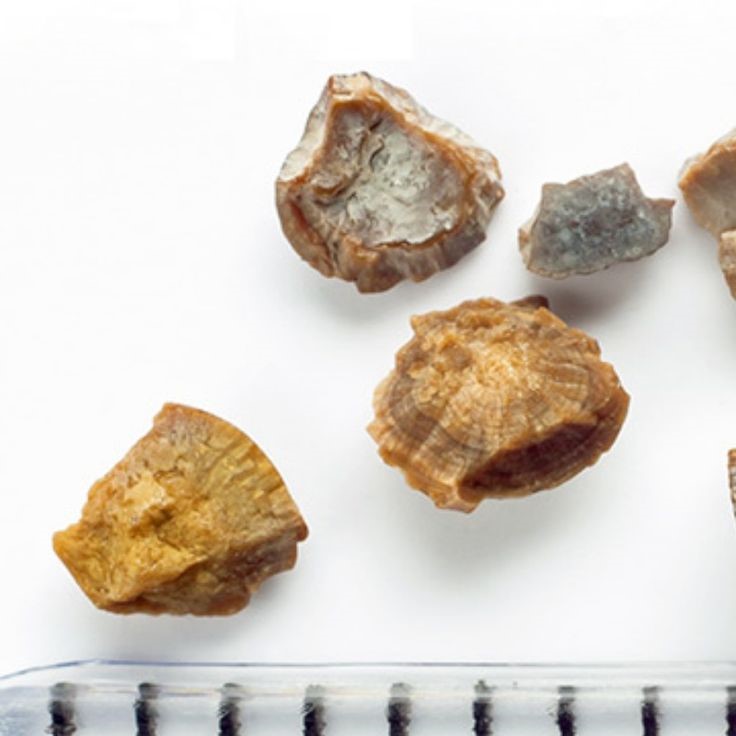
Kidney stones form as hard deposits in the kidneys. They develop from minerals and salts in the urine. Often, they cause pain when passing through the urinary tract. The study of these stones is key to understanding and treating the condition. Analysis often starts with microscopic examination. A kidney stone microscope allows doctors to look at the stones closely. This helps identify the stone’s composition. The type of stone can influence which treatments will work best. For example, calcium stones and uric acid stones require different approaches. Early microscope exams used basic light microscopes. Today, advanced technologies provide clearer images and more detailed information. By using a kidney stone microscope, experts can decide on the most effective treatment. They can also give advice on preventing future stones. Effective analysis leads to better patient outcomes and less discomfort.
The Role of Microscopy in Kidney Stone Examination
The examination of kidney stones greatly relies on detailed microscopy. A kidney stone microscope plays a central role in analyzing these stubborn mineral deposits. It allows medical professionals to see the stone’s texture and structure up close. Identifying the characteristics of the stone is crucial. It can show what materials make up the stone.
Microscopy can reveal if a stone is a calcium oxalate, uric acid, or another type. This insight is vital, as each type responds to different treatments. Thanks to the microscope, doctors can also spot tiny details. These may suggest how the stone formed. They can even show signs of how it might grow. This information is key in crafting a personalized treatment plan.
Not only does microscopy aid in diagnosis, but it also helps in monitoring. Doctors can track the stone’s response to treatment over time. This tracking can inform adjustments for better results. In essence, a kidney stone microscope is an indispensable tool. It ensures accurate diagnosis and guides effective treatment strategies.
Recent Technological Advancements in Microscopic Analysis
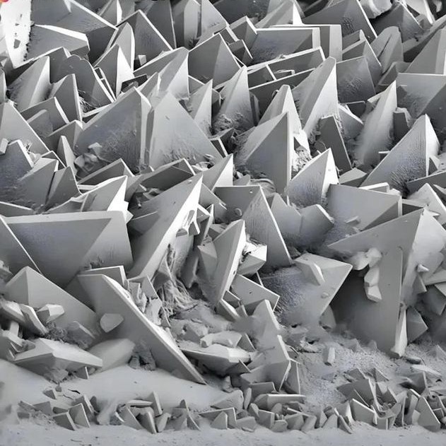
The field of microscopic analysis has seen remarkable growth. Recent advancements have improved our ability to study kidney stones. Modern kidney stone microscopes are now more powerful. They deliver clearer images. High-resolution imaging is a key enhancement. It allows for unparalleled detail during examination. These advances also include better magnification. Doctors can now magnify kidney stones much more without loss of clarity.
Digital microscopy has come to the forefront. It introduces image analysis software. This software helps in quantifying the stone’s components. It also aids in determining their distribution. This level of analysis was not possible before. Another leap in technology is 3D imaging. It gives a better sense of the stone’s shape and features.
Enhanced lighting techniques have emerged as well. They provide more contrast in the images. This makes it easier to identify different types of stones. Each technological step leads to more precise diagnoses. As a result, patients receive more targeted treatments. These advancements also contribute to research. They offer deeper insights into how kidney stones form and grow.
These improvements to the kidney stone microscope have been game-changing. They have vastly enhanced the capabilities of medical professionals. This means better care for those affected by kidney stones. In summary, technology is taking kidney stone analysis to new heights. With it, treatments and patient outcomes improve.

Types of Microscopes Used for Kidney Stone Analysis
When analyzing kidney stones, different types of microscopes are pivotal in yielding accurate results. The type of kidney stone microscope used can vary based on the analysis required. Light microscopy remains one of the most common forms used in initial examinations. It uses visible light to illuminate features of the stones. This type of microscope provides a broad look at stone structure and composition.
Polarizing microscopes are another option. They employ polarized light to highlight the crystalline structure. This method is especially useful for identifying calcium oxalate stones. With polarization, certain materials within the stone can appear more clearly.
Electron microscopes take it a step further with higher resolution. They offer much greater magnification than light microscopes. They allow doctors to see the stone surface at a molecular level. This is critical for identifying very fine details that other microscopes might miss.
Another option includes confocal microscopes. These use laser scanning to produce high-resolution 3D images. They can show the stone’s depth and complexity in detail. This aspect is crucial for understanding the stone’s formation.
Each type of kidney stone microscope has its own strengths. Doctors choose based on what they need to find out. By combining these tools, they can get a complete picture of the stone. This allows for a more informed and effective treatment plan.
How Microscopic Analysis Aids in Treatment Planning
The use of a kidney stone microscope in treatment planning is invaluable. Doctors examine stones with such microscopic tools to decide on the best medical approach. Here’s how microscopic analysis assists in the process:
- Stone Composition Analysis: By examining the stone’s makeup, doctors can prescribe specific dietary changes or medications. For instance, if a kidney stone microscope reveals a high amount of calcium oxalate, treatment may involve a diet low in oxalate-rich foods.
- Treatment Response Monitoring: With ongoing microscopic analysis, practitioners can see how a stone reacts to treatment. They can adjust the plan for better results based on the stone’s changes.
- Surgical Strategy: In cases where surgery is needed, detailed images from the kidney stone microscope guide surgeons. They plan their approach with a clear understanding of the stone’s size and position.
- Prevention Strategies: Understanding a stone’s formation and growth can help prevent new stones. Doctors use this data to advise patients on long-term prevention measures.
Microscopic analysis is a cornerstone of kidney stone management. It allows for personalized and precise treatment strategies. This leads to improved outcomes for patients suffering from kidney stones. Doctors coordinate care with detailed knowledge obtained from microscopic scrutiny. This offering does not just treat but also helps in preventing future stones. In short, a kidney stone microscope is crucial for effective kidney stone treatment planning.

Innovations in Imaging Techniques for Kidney Stone Analysis
In recent years, innovative imaging techniques using a kidney stone microscope have revolutionized the analysis of kidney stones. These advancements facilitate a deeper understanding of stones, aiding in more accurate diagnoses and refined treatment plans. Here are some of the cutting-edge techniques now in use:
- High-Definition Imaging: Today’s kidney stone microscopes offer incredibly high-definition images, exposing minute details of stones that were once impossible to see with older equipment.
- Digital Image Enhancement: Digital tools now enhance microscopic images, providing clearer views and helping to discern the subtle differences between types of kidney stones.
- Software-Assisted Analysis: Sophisticated image analysis software can now measure and analyze the composition and distribution of the materials that make up a kidney stone, giving precise data for treatment decisions.
- 3D Imaging Capabilities: Three-dimensional imaging has taken the stage, allowing doctors to observe the stone’s structure from multiple angles, leading to insights into its formation and potential treatment routes.
- Live Imaging During Procedures: Microscopes equipped with live imaging technology can now be used during procedures, giving surgeons real-time feedback as they work to remove or break down stones.
These innovations are a clear leap forward from traditional methods. They not only improve the examination process but also enhance patient care by contributing to the development of more effective treatments for kidney stones. As the technology continues to advance, we can expect even greater breakthroughs in the microscopic analysis of kidney stones, paving the way for new solutions in the fight against this painful condition.
Comparing Traditional and Modern Microscopic Methods
When discussing the evolution of kidney stone analysis, comparing traditional and modern microscopic methods highlights significant progress. Traditional microscopy offered basic insights into stone composition, often through light microscopes. These instruments provided general information on stone size, shape, and surface details. However, they had limitations. Magnification was lower, and clarity could diminish at higher levels of zoom, making detailed analysis difficult.
In contrast, modern methodologies using a kidney stone microscope are far more sophisticated. They employ advanced technologies that were once unavailable or impractical. High-resolution imaging is now standard, enabling medical professionals to see fine details that inform diagnoses and treatments. Digital microscopy has revolutionized the field as well, allowing for image enhancements and precise measurements. Moreover, with the advent of 3D imaging, doctors gain a comprehensive view of kidney stones, understanding their structure in ways traditional methods couldn’t achieve.

The impact on treatment planning is substantial. More information translates to better-targeted therapies, reducing trial and error in finding effective solutions. Improved imaging allows for closer monitoring of how stones respond to various treatments, leading to timely adjustments and improved patient outcomes.
Overall, the leap from traditional to modern microscopy has resulted in a quantum leap in the quality of care for kidney stone sufferers. Modern tools empower doctors with data that is precise, detailed, and actionable. This technological evolution underscores the critical role microscopy plays in kidney stone analysis and treatment.
Future Directions in Microscopic Analysis of Kidney Stones
The evolution of the kidney stone microscope continues to progress rapidly. As we look ahead, several promising directions suggest how microscopic analysis will advance even further.
Enhanced Resolution and Magnification
- Higher Resolution Capabilities: Future advancements in microscopy technology may allow for significantly higher resolutions, permitting scientists and medical professionals to observe kidney stone structures in greater detail.
- Detailed Structural Analysis: Enhanced magnification would enable a deeper investigation into the crystalline structures and composition of kidney stones, aiding in better understanding and characterization.
- Implications for Research: This level of detail can facilitate innovative research and development of more effective treatment options by revealing previously unseen features of the stones.
Integration of Artificial Intelligence (AI)
- Image Interpretation: Artificial intelligence could be harnessed to analyze microscope images and identify patterns or abnormalities in the kidney stone structures automatically.
- Faster Diagnoses: AI algorithms could significantly speed up the diagnostic process by quickly analyzing images and generating reports, allowing healthcare providers to make informed decisions without delay.
- Predictive Capabilities: By utilizing historical data and advanced algorithms, AI could predict stone behavior, such as likelihood of growth or potential complications, providing valuable insights for treatment planning.
Automated Image Analysis
- Machine Learning Algorithms: The use of machine learning can enable automated analysis of microscopic images, reducing the need for constant human oversight and intervention.
- Efficiency in Results: Automated systems could lead to faster processing times, resulting in quicker turnaround for test results, which is critical in acute medical situations.
- Data-Driven Insights: The analysis could also identify trends or anomalies that might be overlooked by human eyes, further enhancing diagnostic accuracy.
Portable Microscopy Devices
- In-Clinic and At-Home Monitoring: The development of portable microscopes could allow for on-site examinations and easy monitoring of kidney stones both in clinical settings and at home.
- Convenience for Patients: Patients could benefit from more accessible follow-ups and routine checks without the need for extensive hospital visits, leading to enhanced patient compliance and satisfaction.
- Real-Time Monitoring: These devices enable real-time assessment of kidney stone progression, facilitating timely interventions and better management of patient health.
Molecular Imaging Techniques
- Identifying Molecular Makeup: Emerging imaging technologies may allow precise visualization of kidney stones at the molecular level, helping to clarify their composition and structure.
- Early Detection of Stone Formation: Advanced molecular imaging could assist in identifying the initial stages of stone formation, enabling earlier intervention and preventative measures.
- Customized Treatment Approaches: Understanding the molecular characteristics of kidney stones can lead to tailored treatment strategies based on specific stone types.
Nanotechnology Applications
- Enhanced Imaging Techniques: Nanotechnology could refine imaging processes, providing sharper, more detailed images of kidney stones and surrounding tissue.
- Direct Treatment Options: Innovative applications of nanotechnology may lead to techniques that target kidney stones directly, delivering treatments more effectively and potentially minimizing side effects.
- Revolutionizing Kidney Stone Management: The integration of nanotech could transform how kidney stones are diagnosed, treated, and monitored, paving the way for major advancements in urology.
These forward-looking innovations would not only upgrade diagnostic techniques but also pave the way for more personalized and effective treatment strategies. Moving forward, it is clear that the kidney stone microscope will remain an essential tool in battling kidney stone diseases, making significant strides in patient care and quality of life.


