Introduction to Animal Cells
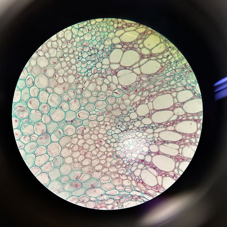
Animal cells are the basic units of life in organisms of the animal kingdom. These cells are incredibly complex, with various structures, known as organelles, each with specific functions. Observing an animal cell under a microscope opens up a fascinating world. It allows us to see components normally invisible to the naked eye. Unlike plant cells, animal cells do not have a rigid cell wall, which gives them a unique, flexible shape. Instead, they are encapsulated by a plasma membrane that controls the entry and exit of substances. Inside, the cell houses organelles such as the nucleus, mitochondria, and endoplasmic reticulum. These structures work together to sustain the cell’s life processes.
Despite their microscopic size, their contribution to life is immense. Studying an animal cell under microscope reveals these components and the vital roles they play. It gives us insights into the complex mechanisms of life, health, and diseases that affect various animal species, including humans. For students, scientists, and curious minds, understanding animal cells is the first step toward unlocking the mysteries of biology.
The Microscope: A Window to Cellular Wonders
The microscope serves as a crucial tool in biology. It allows us to delve into the minute world of animal cells. Through its lenses, we gain the ability to observe cells and organelles. This instrument magnifies the otherwise unseen. It affords us a detailed look at cell structure and function. When we peek through a microscope, we are looking into a world as complex as any we can see with our naked eye. Microscopy has transformed our understanding in fields from medicine to marine biology.
Researchers rely on this tool to study the fine details of animal cells. Students use microscopes to learn about cellular processes from an up-close perspective. In essence, the microscope acts as a bridge. It connects the macroscopic world we live in to the microscopic world of cells. With a good microscope, one can observe the intricacy of an animal cell under microscope. The cell’s dynamic activities become visible, revealing the beauty of biology’s building blocks.
Preparing Animal Cells for Microscopic Examination
Before we can observe an animal cell under a microscope, we need to prepare it correctly. This preparation is vital to ensure we can clearly see the structures within the cell. There are several steps to follow.
- Sample Collection: First, we collect the cells. We can obtain these from tissues or through cell cultures.
- Cleaning: Next, we clean the sample. This removes any debris or substances that might obscure our view.
- Fixation: We then fix the cells. This process preserves their structure. It prevents decay and maintains the cell’s shape.
- Staining: After fixation, staining is often next. Dyes make the cell’s components stand out. This step is crucial because many organelles are nearly invisible without it.
- Mounting: Finally, we mount the cells on a slide. We cover them with a cover slip to protect them and to keep them in place.
Each step is essential for a clear view of an animal cell under a microscope. Proper preparation leads to precise observations. Skipping steps or doing them poorly can ruin a sample. It can also lead to false observations. For anyone studying biology, mastery of these steps is key. It ensures that what we observe through the microscope is an accurate representation of the cell’s structure and function.
Key Structures of Animal Cells Observed Under the Microscope
When examining an animal cell under a microscope, certain structures become visible. These are crucial for the cell’s survival and function. Below are the key structures often observed:
- Nucleus: This is the cell’s command center. It contains DNA, the genetic material that guides cell activities.
- Mitochondria: Often called the ‘powerhouses’ of the cell. They produce energy for the cell through respiration.
- Endoplasmic Reticulum (ER): The ER has two types. The rough ER helps in protein synthesis. The smooth ER manages lipid production and detoxification.
- Golgi Apparatus: This is the packaging and distribution center. It modifies proteins and lipids. Then, it sends them to their destinations.
- Lysosomes: They are the cleaning crew. Lysosomes digest waste and foreign invaders that enter the cell.
- Cytoplasm: This is the jelly-like fluid that fills the cell. It holds all organelles in place.
- Cytoskeleton: This network of fibers gives the cell shape and aids in movement.
- Plasma Membrane: It is the cell’s outer lining. The membrane controls what enters and leaves the cell.
These structures are generally too small to see with the naked eye. But with a microscope, we gain a clear picture of how each works and interacts within the cell. This microscopic view is essential for understanding cell function, health, and disease.
Understanding Cell Division: Mitosis in Animal Cells
Cell division is a vital process for life. In animal cells, this division happens through mitosis. Mitosis leads to growth and repair in multicellular organisms. It ensures that each new cell gets a copy of the genetic material.
Mitosis occurs in five stages: prophase, metaphase, anaphase, telophase, and cytokinesis. During prophase, chromosomes become visible. The nuclear membrane starts to break down. In metaphase, chromosomes line up in the cell’s center.
Anaphase is where the chromosomes split. Each half moves to opposite ends of the cell. Telophase is the reverse of prophase. Here, new nuclear membranes form around the chromosome groups. Finally, cytokinesis divides the cell into two. Each cell has a full set of chromosomes.
Watching mitosis under a microscope is amazing. You can see the chromosomes’ dance during cell division. Observing mitosis helps understand how tissues grow, heal, and maintain their functions. It also sheds light on what goes wrong in diseases like cancer. When cells divide uncontrollably, it can lead to tumors.
To see mitosis, scientists often use cells from an ‘actively dividing’ stage. These come from tissues like bone marrow or root tips. They stain the cells to make the chromosomes stand out. Then, they can track each stage of mitosis.
Understanding mitosis is crucial for biology students and researchers. It is the foundation of cell biology and critical for advanced medical research. Seeing an animal cell under microscope go through mitosis is witnessing life’s complexity in its most fundamental form.
The Role of Stains in Enhancing Microscopic Visibility
Stains play a pivotal role in animal cell microscopy. These special dyes add contrast to the otherwise transparent cellular structures. Without stains, most cell parts would remain invisible even under a microscope. Stains bind to specific components, making them stand out. This improved visibility is crucial for detailed study.
Biologists use various stains depending on the cell parts they wish to observe. For example:
- Hematoxylin and eosin (H&E): This common combination stains cell nuclei blue and other structures pink.
- Methylene blue: Preferred for highlighting DNA in the nucleus.
- Iodine solution: Stains starch in cells, useful for identifying glycogen in animal cells.
Choosing the right stain is vital for accurate observation. Some stains react with specific cell parts only. This specificity helps in identifying various organelles. For instance, the acidic parts of the cell take up basic dyes. Meanwhile, basic parts pick up acidic dyes.
Staining procedures also differ. Some require a single dye, while others need a sequence of stains. The choice of procedure affects the final image. Proper staining can reveal the intricate details of an animal cell under microscope. It can show the nucleus, mitochondria, and even smaller organelles like ribosomes.
In summary, stains are essential tools in microscopy. They provide the contrast needed to see the wonders of animal cells. The right use of stains reveals the dynamic world within cells. It enhances our understanding of cellular structure and function.
Common Observations in Animal Cell Microscopy
When observing an animal cell under microscope, some sights are quite common. These repeated patterns tell us about the cell’s health and activity. Here are some typical observations:
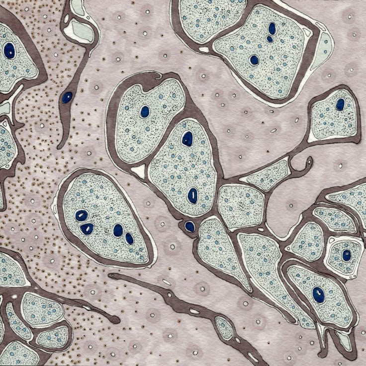
- Cell Shape: Animal cells often appear round or irregular. This shape flexibility helps them to function in varied environments.
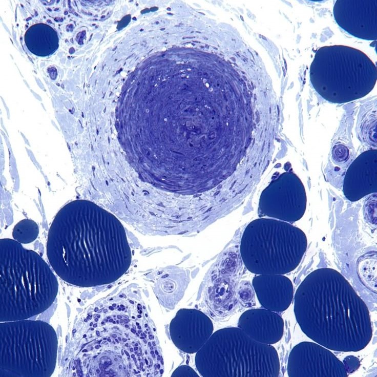
- Nucleus: A dark spot, usually in the cell center, marks the nucleus. It holds the cell’s DNA.
- Cytoplasm: This fluid fills the cell. It appears as a clear area around the nucleus and organelles.
- Cell Membrane: Surrounding the cell, it might show waves or ripples. This indicates interaction with the surrounding environment.
- Mitochondria: These may appear as tiny rods or ovals. They are easier to see when stained.
Students and scientists watch these common occurrences to learn. They are the basics before studying more complex behaviors. An animal cell under microscope provides a window into the life of the cell, showing us signs of how it breathes, eats, and responds to changes. Each observation can lead to deeper inquiries and discoveries in the world of cell biology.
Advances in Microscopic Techniques for Studying Animal Cells
As we explore the microscopic realms, recent advances have enhanced our study of animal cells under microscope. These innovative techniques have improved how we observe and understand cellular details. Here are some significant developments:
High-Resolution Microscopy
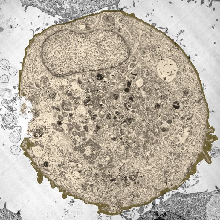
- Advancements in Magnification: Modern microscopes have made significant strides in providing higher magnification and improved resolution. This enhanced capability allows scientists to observe cellular structures with unprecedented clarity.
- Detail Recognition: Thanks to these developments, we can now explore the intricate details of cell parts, such as organelles and membranes, more effectively than ever before. This intricate observation aids researchers in better understanding cellular functions and interactions.
- Applications in Research: High-resolution microscopy is invaluable in various fields including biology, material science, and medicine. Researchers utilize this technology to gain insights into cellular processes, disease mechanisms, and nanotechnology applications.
Fluorescence Microscopy
- Fluorescent Dyes Utilization: Fluorescence microscopy employs special fluorescent dyes that emit visible light when exposed to specific wavelengths of light. This feature highlights certain areas within the cell, making crucial cellular components more visible.
- Specificity in Targeting: By using different fluorescent dyes, researchers can label and visualize specific proteins, nucleic acids, or organelles. This selective visualization is vital for studying the localization, dynamics, and functions of specific cellular components.
- Enhanced Study Opportunities: Fluorescence microscopy opens new avenues for investigating complex cellular processes, such as protein interactions or metabolic pathways. This technique aids greatly in cell biology and pharmacology research.
Confocal Microscopy
- Focused Imaging Techniques: Confocal microscopy improves image clarity by focusing on a single plane within a specimen, allowing for clearer visuals of cellular structures. This technique minimizes the overlapping of light from different layers of the sample.
- Reduction of Blurriness: By scanning through a specimen one plane at a time, confocal microscopy effectively eliminates blurry sections that can occur in traditional imaging techniques. This results in sharper, more detailed images.
- 3D Reconstruction Possibilities: Confocal microscopy is instrumental in creating three-dimensional reconstructions of cells and tissues. By capturing multiple planes, scientists can build a spatial model for better understanding of complex structures.
Live Cell Imaging
- Real-Time Observation: Recent advancements in microscopy have made it possible to visualize living cells over extended periods. This feature allows researchers to see how cells behave and interact in real-time under physiological conditions.
- Dynamic Changes: By examining live cells, scientists can observe cellular movements, how they divide, and their response to external stimuli. This dynamic imaging is crucial for studying developmental biology and pathology.
- Impact on Drug Development: Live cell imaging plays an important role in drug discovery and testing, allowing researchers to monitor cellular responses to new compounds in real-time, leading to a better understanding of therapeutic effects.
Super-Resolution Microscopy
- Breakthrough Technologies: Super-resolution microscopy has achieved significant breakthroughs that surpass the diffraction limit of light. This advancement allows scientists to visualize structures at the nanometer scale, which was previously unattainable with conventional techniques.
- New Dimensions in Imaging: By utilizing innovative approaches, such as STED (Stimulated Emission Depletion) and SIM (Structured Illumination Microscopy), researchers can obtain images with remarkable detail that reveal intricate cellular structures in unprecedented clarity.
- Applications in Nanotechnology: The ability to see at nanometer scales is vital for advancing studies in nanotechnology, enabling more precise manipulation of materials and better understanding of nanoscale biological processes.
Automated Image Analysis
- Software Innovations: The advent of sophisticated software for automated image analysis allows for quicker and more accurate interpretation of cell images. Researchers are now able to process large volumes of data with minimal manual intervention.
- Enhanced Efficiency: By automating the analysis process, scientists save significant time that can be allocated to other important research activities. This efficiency enables faster progress in experimental research.
- Improved Data Interpretation: Automated systems can reduce human error and bias in data interpretation, leading to more reliable results. This improvement is particularly valuable when dealing with complex datasets generated by modern imaging techniques.
Each of these techniques pushes the boundaries of what we can see and learn from an animal cell under microscope. They are vital tools for researchers, offering new insights into cell functions and diseases. Observing animal cells has not only become more precise but also more dynamic, revealing the ongoing dance of life within the confines of a tiny, yet complex, world.


