Introduction
The compound microscope, a cornerstone in scientific laboratories, is a precision instrument. It magnifies tiny objects, enabling us to explore the microscopic world. Each part of a compound microscope plays a crucial role in its functionality. Understanding these parts helps users achieve consistent, clear, and accurate observations. This post delves into compound microscope parts, shedding light on their importance. Stay tuned to uncover the secrets of eyepieces, objective lenses, and more.
Key Components of a Compound Microscope
Understanding the key components of a compound microscope is essential for any user. These parts work together to magnify specimens and reveal details unseen by the naked eye. Let’s take a closer look at the main features that make up the anatomy of a compound microscope.
Optical System: Eyepiece and Objective Lenses
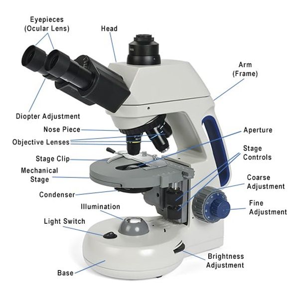
The optical system is at the heart of the microscope’s function. It comprises the eyepiece (or ocular lens) and a series of objective lenses. The eyepiece usually has a magnification power of 10x, bringing the image closer to your eye. Objective lenses, usually found in a rotating nosepiece, come in various magnifications, often ranging from 4x to 100x. Together, eyepiece and objective lenses combine their magnifying powers to create a detailed image of the specimen.
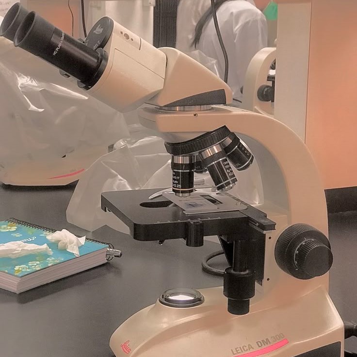
Structural Elements: Base, Arm, and Stage
The structural integrity of a compound microscope rests on its base, arm, and stage. The base provides stability, making sure the instrument does not tip over. Attached to the base, the arm supports the upper parts and allows for easy carrying. The stage holds the slides in place and often includes clips to secure them. It’s a platform for observing specimens and can move to better position the sample under the lenses.
Focusing Mechanism: Coarse and Fine Adjustment Knobs
Every compound microscope has a focusing mechanism, crucial for getting a clear image. It includes coarse and fine adjustment knobs. The coarse adjustment knob makes large movements, bringing the viewer into a rough focus range. The fine adjustment knob is for precision, fine-tuning the focus until the details are sharp and clear.
Illumination System: Light Source and Condenser
For you to see anything under a microscope, you need light. The illumination system includes a light source, often LED or halogen, and a condenser. The condenser focuses the light onto the specimen, ensuring even lighting and improved contrast. This system helps highlight the details of the specimen being observed.
Specimen Handling: Slides and Coverslips
Finally, proper specimen handling is crucial. Slides are thin pieces of glass that hold the sample. Coverslips, which are smaller, thinner pieces of glass, are placed over the sample. They protect the specimen and lens from contamination. Together, slides and coverslips keep the specimen flat and in the correct position for observation.
The Role of Magnification and Resolution
Understanding the role of magnification and resolution is key in using a compound microscope effectively. Magnification makes objects appear larger, while resolution defines the clarity of the image. Together, they affect how we view specimens under the microscope. High magnification with poor resolution can lead to a blurry image. Good resolution means you can distinguish two points as separate. The goal is to achieve a balance between the two for the best image quality.
In compound microscopes, magnification is a product of two systems. The eyepiece, usually at 10x power, works with the objective lenses to enlarge the image. For example, an eyepiece at 10x paired with a 40x objective lens results in a total magnification of 400x. But magnification alone is not enough.
Resolution plays a vital role in bringing out the details. It depends on the quality of the lenses and the wavelength of light used. A higher resolution can separate fine details in the specimen that might otherwise blur together at lower resolution.
The combination of the microscope’s optics (lenses) and proper illumination is crucial for achieving high resolution. Clean, well-maintained compound microscope parts support better imaging results. Users must understand how to adjust both magnification and resolution to examine their samples most effectively.
Care and Maintenance of Compound Microscope Parts
To keep a compound microscope in top condition, proper care and maintenance are crucial. This not only ensures accurate results but also prolongs the life of the instrument. Here are the essential steps for preserving the functionality and clarity of your microscope’s components:
- Handle with Care: Always grasp the arm and base when moving the microscope. Avoid touching the lenses with your fingers.
- Clean Regularly: Use a lens cleaner and a soft, lint-free cloth to gently wipe the optical parts. For the body and stage, a damp cloth will do.
- Check the Illumination: Ensure the light source is functioning correctly. Replace bulbs or LEDs when necessary to maintain consistent lighting.
- Adjustments: After use, turn the coarse and fine adjustment knobs to their neutral positions to reduce tension.
- Storage: Cover the microscope with a dust cover when not in use. Store it in a dry, temperature-controlled environment.
- Regular Inspections: Periodically check all parts, such as the eyepiece, objective lenses, and stage clips, for stability and wear.
These simple actions can safeguard your investment and keep the compound microscope parts in peak condition, ready for your next scientific exploration.
Common Issues and Troubleshooting Tips
When using a compound microscope, users may encounter several common issues. Identifying and resolving these problems is essential for seamless operation. Below are tips for troubleshooting frequent challenges linked with compound microscope parts.
- Blurred Vision: If the image is not clear, check if the lenses are clean. Gently wipe them using lens cleaning solution and a microfiber cloth. Make sure the objective lens is correctly clicked into place.
- Difficulty Focusing: If you can’t get a sharp image, adjust the coarse and fine focus knobs. Start with the coarse knob for general focus and then use the fine knob for detailed clarity.
- Inadequate Illumination: Confirm that the microscope’s light source is working. If not, you might need to replace the bulb or ensure it’s properly connected. Also, adjust the condenser to direct more light onto the specimen.
- Image Distortion: This could be due to incorrect placement of the specimen or coverslip. Ensure the specimen is flat and the coverslip is applied without air bubbles.
- Objective Lens Issues: If an image cannot be brought into focus at different magnifications, ensure that each objective lens is clean and securely mounted in the revolving nosepiece.
- Mechanical Problems: If the stage or knobs are stuck, don’t force them. Check for any obstructions and clean any debris around the moving parts.
Addressing these issues can enhance your experience with compound microscope parts. Routine inspections and maintenance can prevent many of these problems. By applying these tips, you can troubleshoot most common issues efficiently and ensure the microscope functions optimally for accurate scientific investigation.
Innovations and Advances in Microscope Technology
The world of microscopy continues to evolve with remarkable technological advancements. These innovations aim to enhance the capabilities of compound microscope parts, offering better resolution, easier use, and more detailed imaging. Here’s a look at some of the forward strides in microscope technology:
Digital Integration
- Modern compound microscopes are now equipped with digital cameras and software integration, marking a significant advancement in microscopy technology.
- This integration enables users to capture high-quality images of specimens and store them digitally on a computer for later analysis.
- With specialized software, users can enhance, annotate, and organize their images, streamlining both research and educational processes.
- These digital features facilitate remote sharing and collaborations, making it easier for scientists and students to present their findings and insights to a broader audience.
- Additionally, being able to analyze images using digital tools allows for more intricate examinations, leading to improved understanding and discovery.
LED Illumination
- The transition to LED light sources in modern microscopes has provided numerous benefits over traditional halogen bulbs.
- LEDs offer consistent illumination, ensuring that specimens are evenly lit without glare or hotspots, which can hinder visual clarity.
- These light sources are highly energy-efficient, reducing the overall power consumption of the microscope.
- Moreover, LEDs produce significantly less heat compared to halogen bulbs, which helps protect heat-sensitive specimens and maintain a stable working environment.
- Cyclable LED technology also extends the lifespan of the light source, resulting in reduced maintenance costs and improved reliability in long-term usage.
Fluorescence Microscopy
- Advances in fluorescence microscopy have transformed how researchers visualize specific components within cells or microorganisms.
- Modern techniques and new fluorescent dyes allow for the targeted labeling of various cellular structures, enhancing the ability to study complex biological processes.
- With improved clarity and specificity, fluorescence microscopy enables scientists to observe interactions within cells in real time, advancing fields such as cellular biology and pathology.
- The ability to apply multiple fluorescent markers simultaneously opens up new avenues for multi-parameter analysis of cellular functions.
- As a result, researchers can gather more comprehensive data, which aids in deeper understanding and potential breakthroughs in medical research.
Phase Contrast and DIC
- Phase contrast microscopy and Differential Interference Contrast (DIC) are innovative techniques that enhance the visibility of unstained samples.
- These methods improve contrast through optical phase shifts, allowing for visualization of fine details without the need for dyes or stains, which can sometimes alter sample characteristics.
- Phase contrast microscopy is especially useful for observing living cells, as it enables real-time analysis of cell behavior in their natural state.
- Similarly, DIC produces images with a three-dimensional quality, enhancing the perception of depth and texture in specimens.
- Both techniques provide researchers with powerful tools for studying samples that are otherwise difficult to visualize, leading to more accurate observations and analyses.
Automation
- The incorporation of automation into microscopy has significantly enhanced efficiency and precision in scientific research.
- Automated stages and focusing systems enable smooth and reproducible movements, reducing the risk of human error during sample positioning and focusing.
- With automated setups, users can easily program the microscope for various tasks, such as scanning large samples or conducting time-lapse studies without manual intervention.
- This feature saves valuable time in experimental setups and allows for more extensive data collection without sacrificing quality or accuracy.
- By minimizing manual handling, automated systems also reduce the likelihood of sample contamination and ensure consistency in results.
Improved Objective Lenses
- Advances in objective lens technology continue to redefine the capabilities of modern microscopes, pushing the limits of resolution and magnification.
- New lens designs incorporate sophisticated coatings and materials that minimize optical aberrations, leading to sharper and clearer images.
- These improvements enhance the overall image quality, allowing for detailed observations even at high magnifications.
- Cutting-edge lens technologies enable better light transmission, which is essential for observing faint specimens or fluorescent markers effectively.
- As resolution and clarity improve, researchers can gain finer insights into microstructures, paving the way for discoveries in various scientific fields, including materials science, biology, and nanotechnology.
These developments represent just a glimpse into the continuous improvements being made in the field of microscopy. Through these innovations, compound microscopes provide scientists and researchers with an unprecedented view of the microcosm, fueling discovery and increasing our understanding of the world at the microscopic level.
Conclusion: Enhancing Scientific Discovery with Compound Microscopes
In the quest for scientific discovery, compound microscopes play a key role. They let us see beyond the limits of our natural vision. By understanding compound microscope parts and their function, we unlock the potential to observe and analyze the microcosm. Properly using these tools helps us magnify minuscule details and enhance image resolution. This balance is vital for crisp, clear results.
Maintenance is crucial for long-lasting use. Regular care prevents common issues that could blur or distort images. As our guide shows, simple checks and cleanings make a huge difference. Updated technology further boosts compound microscopes’ powers. Digital features and better lenses refine how we explore and document the microscopic world.
As technologies advance, so do our methods. We’re now capturing and sharing scientific findings with ease. We’re also ensuring that these instruments serve us well into the future. Thanks to these innovations, our journey into microscopy will continue to uncover the wonders of the unseen world. As users and enthusiasts, this makes our adventures in microscopy not only possible but more exciting than ever.


