Introduction to Microscope Components
A microscope is an essential tool for scientific discovery. Its various parts work together to magnify small objects. Understanding these components is key when using or studying microscopes. Let’s break down the main parts you’ll find in a microscope diagram labeled.
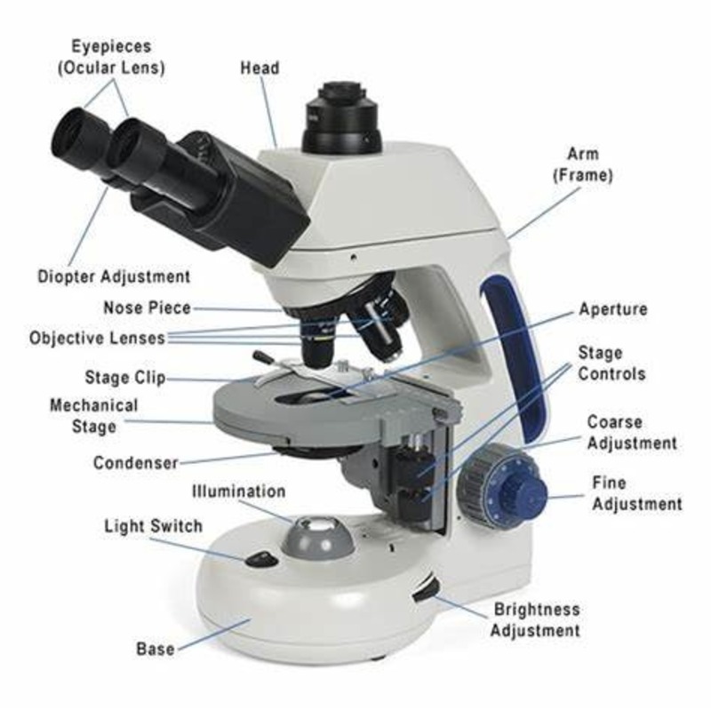
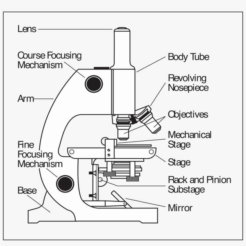
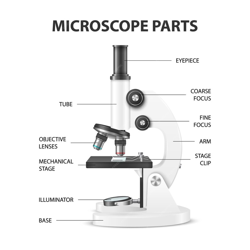
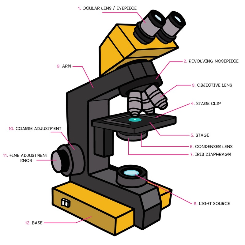
First, there’s the eyepiece lens, also known as the ocular lens. It’s the part you look through. Eyepieces usually provide the first level of magnification for a sample. Then come the objective lenses. These are on a rotating turret and move closer to the specimen for increased magnification.
The stage holds the specimen in place. It typically includes clips to secure slides. Under the stage lies the illuminator. It’s the light source that brightens the object being viewed. Above it, there’s the condenser. It focuses the light onto the sample. Close by is the diaphragm or iris. This part adjusts the amount of light reaching the specimen.
Another critical component is the focusing system, including the coarse and fine focusing knobs. These dials adjust the focus by moving the stage or the objective lenses up and down. Together, all these parts make up the basic structure of most microscopes.
In the following sections, we will explore different types of microscopes. They each have their unique diagram and features to consider. So, keep these basic components in mind as we dive deeper into the world of microscopes. With a microscope diagram labeled, you can identify and understand the role of each part.
Types of Microscopes and Their Diagrams
Understanding the structure of microscopes is easier with a labeled microscope diagram. Different types of microscopes have unique features that cater to their specific uses in science. Let’s explore the diagrams of some common microscopes.
Compound Microscope Diagram
A compound microscope is the standard type used in many labs. It has multiple objective lenses on a rotating turret. This microscope diagram labeled will show those lenses, the eyepiece, stage, and focusing knobs. Users can switch between objectives for varied magnification levels. The light source and condenser are below the stage in these diagrams.
Stereo Microscope Diagram
Stereo microscopes, often used for dissection, show in their diagrams two eyepieces for depth perception. They usually have a lower magnification range. The labeled diagram will highlight the three-dimensional viewing capabilities with dual optical paths for each eye.
Digital Microscope Diagram
Digital microscopes integrate cameras for viewing specimens on screens. In a digital microscope diagram labeled, you’d note the absence of an eyepiece. Instead, it includes a digital sensor and often, built-in illumination systems. These diagrams may also feature software components for image analysis.
Electron Microscope Diagram
The most advanced, electron microscopes use electron beams for imaging. Labeled diagrams show the complex vacuum system, electron gun, and magnetic lenses for focusing the electron beam. They offer much higher magnification, capturing details at the molecular level. These diagrams will also display detectors that capture the interactions between the electrons and the specimen.
Each type of microscope has a distinct diagram labeled to represent its unique structure and functions. It’s crucial to understand the differences to utilize each microscope properly for scientific observations.

Understanding the Magnification System
When using a microscope, magnification is a key factor in viewing tiny details. The magnification system has two main parts: the objective lenses and the eyepiece lenses.
Objective Lenses
Objective lenses are the heart of a microscope’s magnifying power. Each lens has its own level of magnification, indicated by numbers like 4x, 10x, or 40x. The lower magnification lenses provide a wider field of view, while higher ones allow you to see finer details. You turn the rotating turret to switch between different objective lenses, combining their magnification with that of the eyepiece.
Eyepiece Lenses
The eyepiece, or ocular lens, further magnifies the image processed by the objective lenses. Most eyepieces magnify images by 10x. When you look through the eyepiece, you see the specimen magnified by both the objective and eyepiece lenses. This dual magnification system allows microscopes to reveal minute details that are otherwise invisible to the naked eye.
Together, the objective and eyepiece lenses create a powerful magnification system. This system enables scientists and students alike to explore the microscopic world in great detail. A labeled microscope diagram helps in identifying these lenses and understanding their functions. It’s essential for anyone studying or working with microscopes to be familiar with this aspect of microscope anatomy.
Illumination and Focusing Systems
Illumination and focusing systems are fundamental in microscopes. They ensure that you can see specimens clearly.
Light Source
Every microscope needs a good light source. For most models, this is called the illuminator. It shines light from under the stage to light up your specimen. In some microscopes, like compound ones, it’s often a bright, LED or halogen bulb. The light makes it possible for you to see the sample.
Condenser and Diaphragm
Just above the illuminator, you have the condenser. This part focuses the light on your specimen. Right next to it is the diaphragm or iris. You use this to control how much light gets to the specimen. This helps improve the contrast and clarity of the image you see.
Focusing Knobs
There are two main knobs for focusing in most microscopes: the coarse and fine focusing knobs. You use the coarse knob to move the stage up and down for general focus. The fine knob helps you sharpen the image. This way, you can see tiny details on the specimen with higher precision.

Specimen Stage and Clips
The specimen stage is a flat platform that holds the sample under investigation. It’s where you place your microscope slides so you can view them through the lenses. The stage often includes clips, or stage clips, that keep the slide in place during observation. Without these clips, the slide could move and disrupt the focus, especially at higher magnifications.
In some microscopes, the specimen stage is also equipped with a mechanical stage. This feature allows precise movement of the slide using knobs. By turning these knobs, you can move the slide left, right, up, and down very smoothly. This is crucial when you’re trying to examine specific parts of your sample.
Furthermore, the stage might have a hole or opening in the center. This opening allows light from the illuminator to pass through the specimen for better viewing. It’s an essential part of the illumination system, playing a pivotal role in the clarity of the image you see.
In summary, knowing the layout and functionalities of the specimen stage and clips is vital for proper microscope use. They are simple yet integral components, keeping your specimen stationary and centered over the light source for optimal examination. A microscope diagram labeled helps in pinpointing these parts and understanding their importance.
Maintenance and Handling of Microscopes
Proper maintenance and handling are crucial for the longevity and accuracy of microscopes. To ensure that your microscope continues to function effectively, here are some essential tips to follow:
Regular Cleaning
Regular cleaning is vital. Use a soft, lint-free cloth to gently wipe the exterior parts. Clean the lenses with a special lens cleaning solution and tissue to avoid scratches and remove oily residues. Dust off the stage and clips to maintain a clear view. Always remove slides and cover the microscope after use to prevent dust accumulation.
Careful Handling
Always carry your microscope with both hands – one on the arm and the other supporting the base. Avoid touching the lenses with your fingers to prevent smudges and potential damage. When adjusting the focus, turn the knobs delicately to prevent harm to the lenses or slides.
Proper Storage
Store your microscope in a dry, cool place to protect it from moisture and heat. If possible, use a dust cover or place it in a cabinet. Ensure the cords are neatly arranged and the voltage is appropriate for the microscope’s light source to prevent electrical issues.
Regular Inspections
Check your microscope periodically for any signs of wear or damage. Ensure that moving parts like the focusing knobs and rotating turret are functioning smoothly. If you encounter issues, consult a professional for repairs instead of attempting to fix them yourself.
By adhering to these maintenance and handling guidelines, you can keep your microscope in top condition. This not only ensures clarity and precision in your observations but also extends the lifespan of the microscope. Always have a microscope diagram labeled handy, as it can assist you in identifying parts that may need attention or replacement.

Conclusion: The Significance of a Labeled Microscope Diagram
A labeled microscope diagram is very important for anyone using a microscope. It helps you identify and understand each part. This makes it easier to use the microscope correctly. Understanding the diagram helps in learning how to prepare slides. It also aids in adjusting the magnification and focusing on the specimen.
For students and new scientists, a labeled diagram is a learning tool. It is a guide for understanding complex scientific equipment. For teachers, it is useful in explaining the functions of each part. This helps students grasp microscope concepts better.
In practical uses, referring to a diagram ensures correct maintenance practices. You know where to clean and how to handle each part. It helps you store the microscope properly to keep it in good shape.
Above all, a labeled diagram enhances safety. Users avoid damaging sensitive parts, like lenses and illumination systems. Through understanding the diagram, one can troubleshoot common issues. You can identify problems early and seek help when needed.
In conclusion, a microscope diagram labeled is a key piece of knowledge. It serves many purposes, from education to equipment care. It ensures precise and effective microscopic analysis. This supports scientific discovery and education in many fields.


