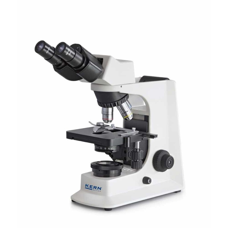
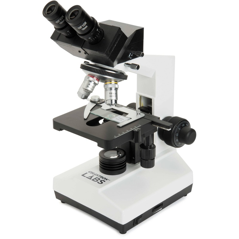
Introduction to Compound Light Microscopes
The compound light microscope is a vital tool in the world of science. It magnifies tiny objects. This helps us see details invisible to the naked eye. Its design uses multiple lenses. The primary lenses are the ocular and the objective ones. First, light passes through the sample. Then it goes through the lenses, creating an enlarged image. The microscope has evolved over centuries. Now, it is a key part of labs across the globe.Explore the anatomy of a compound light microscope labeled diagram. Learn about each part’s function and tips for effective use.
Scientists and students alike rely on this tool. They use it to explore microorganisms, cells, and tissues. A compound light microscope labeled clearly is critical for effective learning. This guide aims to clarify each part’s function. We’ll cover the key components and show you how to use them effectively. By the end of this guide, you’ll understand how the microscope operates. You’ll also get tips on maintenance and troubleshooting. Plus, there’s insight into modern advancements in microscope technology. Let’s dive into the world of the compound light microscope and its wonders.
Key Components of a Compound Light Microscope
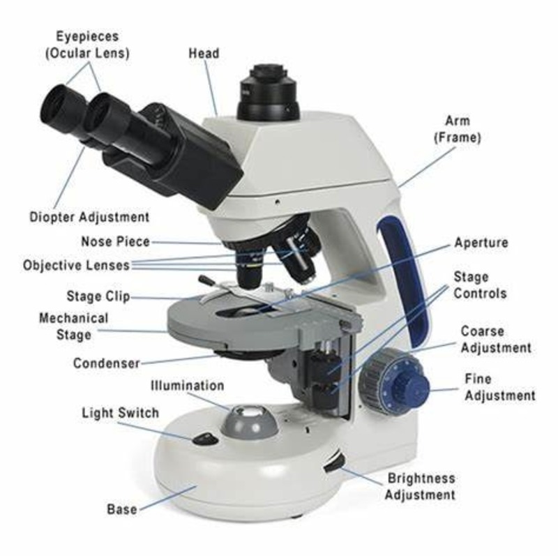
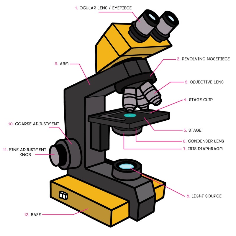
Understanding the various parts of a compound light microscope is crucial for using it correctly. The microscope comprises several key components, each with a distinct purpose. We will explore the eyepiece, objective lenses, stage, and illuminator. Each part plays a role in the microscope’s ability to magnify tiny objects.
The Eyepiece or Ocular Lens
The eyepiece, also known as the ocular lens, is where you look through to see the magnified image. It further enlarges the image formed by the objective lens. Most eyepieces have a magnification power of 10x. This means they make the sample appear 10 times larger than its actual size. When combined with the objective lenses, the final magnification can be quite significant.
The Objective Lenses
The objective lenses are the main magnifiers on a compound light microscope. Usually, there are several, ranging from low to high power. Each lens has different magnifications, commonly 4x, 10x, 40x, and 100x. By rotating the nosepiece, you can switch between these lenses. This allows for various levels of detail to be observed.
The Stage
The stage is the flat platform where you place the slides. It often includes clips to hold your slides in place. The stage can move up or down, allowing focus adjustments. A well-positioned stage ensures that the sample comes into the proper view and focus for observation.
The Illuminator
The illuminator is the microscope’s light source. It shines light up from beneath the stage through the sample. This makes the sample’s details visible. It’s a vital component because without enough light, you cannot see the specimen clearly. Some microscopes have adjustable illuminators, which help control the light’s intensity during observation.
Understanding Magnification and Resolution
To effectively use a compound light microscope, one must grasp the concepts of magnification and resolution. These two factors determine how clear the microscopic image will be. Understanding these principles is key to interpreting what is seen under the microscope.
How Magnification Works
Magnification is the process that makes small objects appear larger. In a compound light microscope labeled with various parts, magnification involves two types of lenses: the ocular and the objective lenses. Here’s how it works: the objective lens first magnifies the specimen. Then, the ocular lens further enlarges this image. The total magnification is calculated by multiplying the magnifying power of the objective lens by that of the eyepiece. So, if you’re using a 40x objective lens with a 10x eyepiece, the image will be magnified 400 times.
Quality microscopes offer a range of objective lenses. These permit viewing at different magnifications. Rotating the nosepiece allows for a seamless switch between these objective lenses.
The Role of Resolution in Clarity
Resolution, or resolving power, is about detail. It’s the capacity of a microscope to distinguish between two close points as separate entities. Higher resolution gives a clearer, more defined image. Achieving high resolution is crucial when magnifying. If the magnification is high but the resolution is low, the image may be large but blurry.
Compound light microscopes rely on good illumination to enhance resolution. The illuminator below the stage plays a vital role in this. Fine adjustment knobs also help in refining the focus, thereby improving resolution. To maximize clarity, the specimen must be properly prepared and the lenses must be clean. These factors work together to ensure that the utmost detail is visible at high magnification levels.
How to Use a Compound Light Microscope Properly
Using a compound light microscope labeled with key components accurately is crucial. It allows precise magnification and observation of microscopic samples. Here’s a simple step-by-step guide to ensure proper usage.
Step-by-Step Usage Guide
- Turn on the illuminator to provide light beneath the stage.
- Place a prepared slide onto the stage using the clips.
- Start with the lowest power objective lens. Rotate the nosepiece to select it.
- Look through the eyepiece and use the coarse adjustment knob to bring the slide into focus.
- Refine the focus using the fine adjustment knob, for a sharp image.
- To increase magnification, switch to a higher power objective lens.
- Adjust the light intensity if necessary, to see the sample clearly.
- Make fine focus adjustments each time magnification changes.
- When finished, lower the stage, remove the slide, and turn off the illuminator.
Remember, starting with the lowest magnification helps to locate the sample. It also reduces the risk of damaging the slide or lenses.
Preparing Slides for Observation
The correct preparation of slides is essential for successful microscopy. Here’s what to do:
- Clean the glass slide and cover slip thoroughly.
- Place the specimen on the slide using tweezers or a needle.
- Add a drop of water or stain if necessary, to enhance visibility.
- Gently lower the cover slip at an angle to avoid air bubbles.
- Dry excess liquid from the slide edges before placing it on the microscope stage.
Proper slide preparation ensures that samples are displayed at their best under the microscope. It helps achieve high-resolution images during observation using your compound light microscope labeled with precision.
Maintaining Your Compound Light Microscope
Proper maintenance of your compound light microscope labeled with precision is crucial. It ensures longevity and reliability. Here, we’ll provide tips for cleaning and storing your microscope. We’ll also guide you through some common issues you might encounter.
Cleaning and Storage Tips
- Use a soft, lint-free cloth to wipe down the microscope body. Do this gently to avoid scratching.
- Carefully clean the lenses with lens paper. Avoid using regular paper or fabric.
- Remove dust from hard-to-reach areas with compressed air.
- Keep the microscope covered when not in use. This protects it from dust.
- Store in a cool, dry place. Extreme temperatures and moisture can damage the microscope.
- Do not disassemble the lenses or mechanical parts unless necessary.
Following these tips will keep your compound light microscope labeled and ready for use.
Troubleshooting Common Issues
- Image is blurry: Check if the lenses are clean. Adjust the focus knobs.
- Light is too dim: Ensure the illuminator is working. Adjust the light intensity.
- Cannot center the specimen: Adjust the slide on the stage properly. Use the stage clips.
- Eyepiece lens is loose: Tighten the eyepiece securely.
- Objectives do not rotate smoothly: Clean the nosepiece. Apply appropriate lubricant if necessary.
With these troubleshooting steps, you can address common issues that affect your compound light microscope labeled components. This will help maintain clear imaging and functionality.
Applications of Compound Light Microscopes
Compound light microscopes labeled with clarity have vast applications. They’re central in fields like education and research, as well as medical diagnostics. Their role is critical for exploring life’s tiny building blocks.
In Education and Research
In academic settings, compound light microscopes serve as hands-on tools for learning. They allow students to explore cell structures and microorganisms first-hand. This practical experience is vital for a solid scientific understanding. For researchers, such microscopes are gateways to new discoveries. They help in observing reactions at the cellular level and understanding disease mechanics.
Scientific publications often include images taken through these microscopes. They document findings crucial for scientific progress. Universities and research institutes invest in high-quality compound light microscopes. They need them labeled and ready for student training and experimental research. Learners get to label parts during lab sessions. This enhances their technical skills and furthers their knowledge.
In Medical Diagnostics
Medical labs use compound light microscopes for diagnosing diseases. Technicians examine blood samples, tissue sections, and other body fluids. They look for abnormalities and infectious agents. This is vital for patient diagnosis and treatment plans.
In pathology, these microscopes allow for detailed examination of biopsied tissue. Doctors can spot cancer cells and other diseases quickly. Using a compound light microscope labeled correctly speeds up this process. It enables precise identification of structures, saving precious time during critical diagnoses.
Their application extends to microbiology labs as well. Here, microbiologists identify bacteria and viruses. They rely on the precise labeling of microscope components for accuracy. Their work is essential in controlling the spread of infectious diseases.
Advances and Innovations in Microscope Technology
The world of microscopy is not static. It keeps evolving with new technologies. Advances in microscope technology have revolutionized what we can see and do. Precise improvements have turned simple models into complex instruments. These advances include better resolution and user-friendly digital features.
Digital Integration and Enhanced Imaging
Modern compound light microscopes labeled with digital technologies are now standard. They blend traditional optics with digital cameras and software. This allows users to capture images directly from the microscope. One can edit, share, and analyze these images using computers. Enhanced imaging features like fluorescence and phase-contrast have also improved. They help in visualizing samples with greater detail.
Digital slides are an innovation worth noting. They let you view samples on computer screens. This is handy for teaching and collaborative research. With image analysis software, one can measure and quantify features easily. Thus, digital integration has made microscopes more versatile than ever.
Future Trends in Microscopy
Looking ahead, the trend is toward more integration and automation. Artificial intelligence (AI) is set to play a bigger role. AI could help with image analysis and even in diagnosing diseases. We might also see improvements in 3D imaging, giving deeper insights into samples.
Additionally, portability is becoming important. Portable light microscopes could bring lab-grade observations to any location. This could be a game-changer for fieldwork or remote diagnostics. We are also moving towards more eco-friendly designs. These microscopes could use less energy and have a smaller environmental impact.
In conclusion, the compound light microscope labeled today could go far beyond its current capabilities. These innovations will continue to open new paths for discovery and learning in various fields.


