Introduction to Hyaline Cartilage
Hyaline cartilage plays a pivotal role in the human body. It is a type of connective tissue prominent in areas requiring support and flexibility. Commonly found in the respiratory tract, the end of long bones, and the connecting surfaces of ribs, its glass-like appearance becomes evident under a microscope. This particular kind of cartilage contains a rich, extracellular matrix that buffers joints and reduces friction.
The fascination with hyaline cartilage under the microscope stems from its complex structure and function. It’s resilient yet supple, making it integral for normal joint movement. Studying hyaline cartilage at the microscopic level reveals insights into its maintenance, repair, and regeneration processes. Researchers rely on the detailed views provided by various microscopic techniques to understand how hyaline cartilage sustains its crucial role in our bodies. This blog delves into the microscopic world of hyaline cartilage, exploring its intricacies and the methods that allow us to visualize its structure with clarity.
The Microscopic Structure of Hyaline Cartilage
Under the microscope, hyaline cartilage reveals an intricate structure. This section focuses on the microscopic architecture, chiefly, the extracellular matrix and chondrocytes.
The Extracellular Matrix
The extracellular matrix (ECM) is a dense network inside hyaline cartilage. It consists of water, collagen fibers, and proteoglycans. Primarily, it provides the cartilage with strength and elasticity. When viewing hyaline cartilage under microscope, the ECM’s role in supporting chondrocytes becomes apparent. Its composition allows for shock absorption, essential in protecting joint integrity.
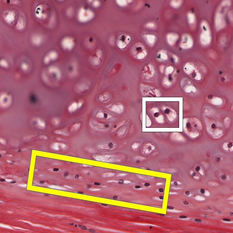
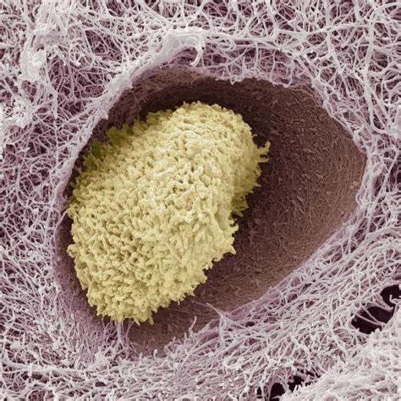
Chondrocytes and Their Lacunae
Chondrocytes are the cells that produce the ECM of hyaline cartilage. Each chondrocyte resides in a small space called a lacuna. These lacunae become visible under magnification. They are crucial for maintaining the cartilage’s health and overall function. Through microscopic examination, one can observe chondrocytes’ involvement in cartilage growth and repair processes.
Methods for Viewing Hyaline Cartilage Under the Microscope
To study hyaline cartilage under microscope, researchers use specific methods. These enhance the visibility of cartilage’s structure and its elements.
Staining Techniques for Cartilage
Staining is vital for examining hyaline cartilage. Stains like hematoxylin and eosin (H&E) enhance contrast. They make the extracellular matrix and chondrocytes stand out. Another common stain, safranin O, highlights proteoglycans. This shows cartilage’s health and progression of diseases like osteoarthritis. Researchers choose stains based on the structure they want to highlight.
Types of Microscopes Used
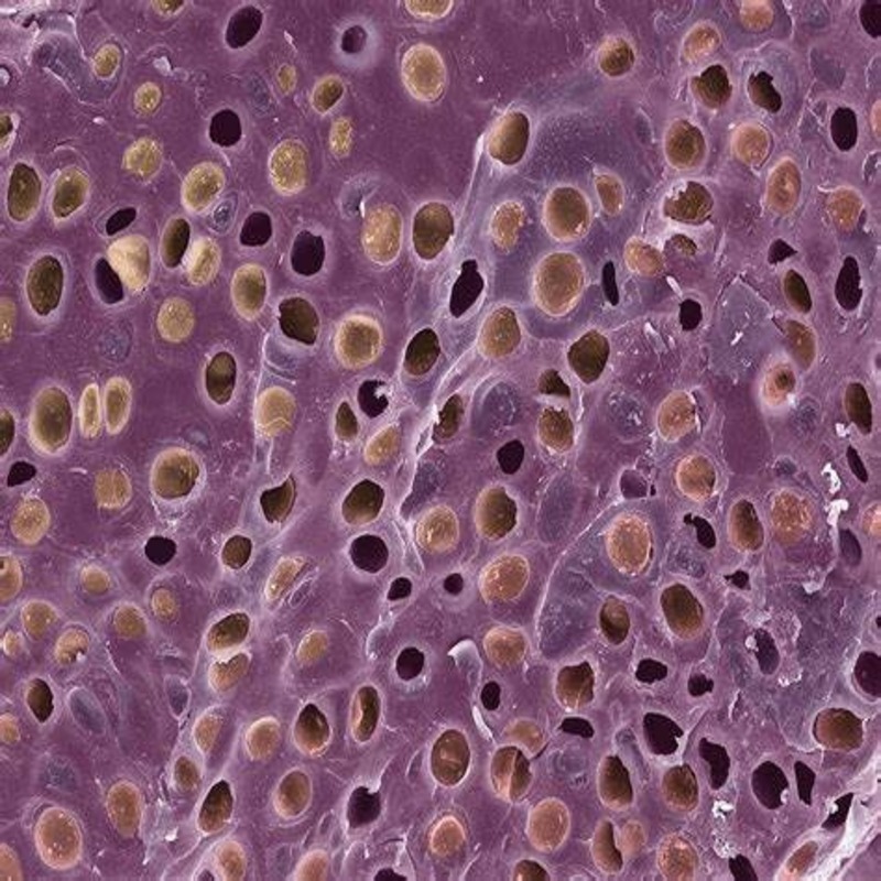
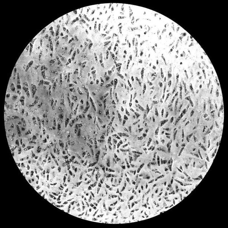
Different microscopes offer unique insights. Light microscopes allow basic viewing of stained cartilage samples. Electron microscopes reveal finer details due to higher magnification. Confocal microscopes enable three-dimensional imaging. This helps in understanding the spatial relationships within hyaline cartilage. Each type of microscope provides valuable information on cartilage structure and function.
The Role of Hyaline Cartilage in Joints
Hyaline cartilage serves an essential function in joint anatomy. It forms what is known as articular cartilage. This smooth tissue lines the ends of bones where they meet to form a joint. Its primary role is to provide a low-friction surface. This allows bones to glide over each other with ease during movement. The quality of hyaline cartilage under microscope is crucial. It shows how well it can absorb shock and distribute loads across the joint. This reduces the risk of bone damage from impact and stress.
Articular Cartilage Function
Articular cartilage functions as a shock absorber in joints. It enables smooth and pain-free movement. When viewed under a microscope, the unique properties of hyaline cartilage come into focus. Its shiny, glassy appearance is due to its smooth surface. This minimizes wear and tear on the joint over time. It has neither blood vessels nor nerves. This feature makes it uniquely suited to function in high-stress environments like knee and hip joints. The resilience of hyaline cartilage is a testament to its role in joint health and mobility.
Hyaline Cartilage Compared to Other Types of Cartilage
In the human body, hyaline cartilage is just one of several types of cartilage. Each type has unique properties and functions. In this section, we’ll compare hyaline cartilage to the other main forms: elastic and fibrocartilage.
Elastic Cartilage
Elastic cartilage is similar to hyaline cartilage but contains more elastin fibers. This composition gives it more flexibility. It’s found in structures needing both shape and bend, like the ear and epiglottis. Under the microscope, we see a dense network of fibers giving rise to its springy texture. Its resilience allows it to withstand repeated bending and returning to the original shape.
Fibrocartilage
Fibrocartilage, on the other hand, is the toughest cartilage type. It has thick bundles of collagen fibers. This structure makes it extremely strong and durable. Fibrocartilage is present in areas with high pressure like intervertebral discs and menisci in knees. Microscopically, its dense and irregular pattern of collagen stands out. It’s this tough matrix that provides unmatched support in high-stress areas.
By comparing hyaline cartilage under microscope with these types, we gain a better understanding of the specialized functions each serves in the body. While hyaline cartilage focuses on flexibility and smooth movement, elastic and fibrocartilage accommodate other needs like shape retention and high-stress support.
Diseases and Disorders Affecting Hyaline Cartilage
Hyaline cartilage, while durable, is not impervious to damage. Certain diseases and disorders uniquely affect this type of cartilage. Understanding these conditions helps in grasping the challenges faced in maintaining joint health. Disorders of hyaline cartilage can lead to pain and reduce mobility. Early diagnosis and treatment are crucial for managing these diseases.
Osteoarthritis and Cartilage Degradation
Osteoarthritis (OA) is a common disorder involving cartilage wear and tear. It typically occurs in joints like the knee and hip. In OA, the smooth surface of hyaline cartilage breaks down. This leads to stiffness, pain, and decreased joint function. Observing hyaline cartilage under a microscope during OA progression reveals a rougher, less uniform surface. The key features include thinning of cartilage and loss of ECM’s cushioning effect.
Chondrodysplasias
Chondrodysplasias are a group of disorders that affect cartilage development. They can lead to abnormal bone growth and joint deformities. These conditions are often genetic in nature. Under the microscope, changes in the structure of hyaline cartilage become visible early on. This includes irregularities in chondrocytes and disruptions in the ECM. Such insights are vital for understanding the disorder’s progression and potential treatments.
Hyaline Cartilage Healing and Regeneration
When injured or damaged, hyaline cartilage faces challenges in healing and regenerating. Unlike other tissues that have a rich blood supply, cartilage is avascular. This means it lacks blood vessels, making nutrient delivery and waste removal inefficient. Moreover, chondrocytes, the cells responsible for cartilage maintenance, have limited ability to replicate and migrate to the damage site. These factors contribute to the slow healing process of hyaline cartilage.
Challenges and Repair Strategies
Rejuvenating damaged hyaline cartilage involves overcoming several obstacles. Key challenges include the avascular nature of cartilage, slow cellular turnover, and intricate extracellular matrix composition. Repair strategies aim to stimulate the regrowth of cartilage and restore joint function.
One approach is using autologous chondrocyte implantation (ACI). This technique harvests chondrocytes from a non-weight-bearing area of the joint. The cells are then cultured in a lab and reimplanted into the damaged site. Another method is microfracture surgery. Surgeons create tiny fractures in the bone beneath the damaged cartilage. This promotes a healing response by stimulating blood flow to the area.
Researchers are exploring tissue engineering as a promising avenue for hyaline cartilage repair. This involves constructing three-dimensional scaffolds to support the growth of new tissue. Advances in biomaterials and growth factors also contribute to developing more effective repair strategies. Combining these with insights from hyaline cartilage under microscope studies will improve treatment methods.
Advancements in Cartilage Research and Microscopy
The field of cartilage research constantly evolves. Recently, advances in microscopy have significantly impacted our understanding of hyaline cartilage. These strides offer new insights into cartilage function and disorders, informing better treatment strategies.
Imaging Technologies and their Impact on Research
Modern imaging technologies have revolutionized the study of hyaline cartilage under the microscope. These improvements widen the scope of what is possible in research. From enhanced resolution to more powerful magnification, the limits of what we can see, and therefore understand, are continually expanding.
Newer imaging tools aid researchers in different ways. They let us see the minute details of cartilage structure. This includes the ECM’s fine makeup and the complex environment of chondrocytes within their lacunae. In diseases like osteoarthritis, these technologies reveal cartilage wear patterns and degradation levels.
One of the standout advancements is the development of high-resolution imaging methods such as multiphoton microscopy. This technique allows for non-invasive, deep-tissue imaging. It helps visualize living tissues in real-time, without the need for traditional staining methods. This is especially crucial for observing processes like cartilage repair and regeneration.
Additionally, advanced imaging software helps quantify changes in cartilage. This can guide the success rate of new treatments. With 3D imaging, we now understand cartilage structure in three dimensions. This depth perception is critical for examining the intricate layering of the ECM.
Overall, these advances enhance our ability to observe hyaline cartilage under a microscope. They grant researchers a detailed view previously not possible. We now look forward to even more sophisticated microscopy techniques. These will continue to illuminate the unseen world of hyaline cartilage and its vital role in joint function and health.


