Introduction to Blood Cell Microscopy
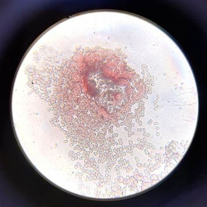
Blood cell microscopy is a fascinating and crucial field in medical science. It involves viewing blood cells under a microscope to diagnose and monitor various health conditions. When we look at blood cells under microscope, we often see a hidden world, teeming with activity. This process is not just about magnifying cells; it is about understanding the life that flows within us.
To begin with, the term ‘microscopy’ refers to the use of a microscope for detailed observation. In the context of blood cells, it entails the use of optical or electron microscopes. These instruments allow scientists and doctors to see structures that are too small for the naked eye. By studying blood cells under microscope, professionals can examine cell shapes, count, and even notice subtle abnormalities that could point to underlying health issues.
The importance of blood cell microscopy lies in its ability to provide valuable insights into an individual’s health. It helps in identifying diseases like anemia, infections, clotting disorders, and blood cancers. Through this method, it is possible to detect changes that might not be evident through regular examinations.
Analyzing blood cells under microscope requires precision and skill. One must carefully prepare the blood sample, stain it to enhance visibility of the cells, and then place it under the microscope for observation. Technicians and pathologists look for any irregularities in size, shape, and number of blood cells, which could be early indicators of diseases.
Understanding blood cell microscopy is crucial for anyone interested in the medical field, especially those specializing in hematology. It provides a window into the complex systems that keep us alive. As the technology improves, so does our ability to diagnose and treat various blood-related conditions more effectively.
The Components of Blood and Their Functions
Blood, a vital fluid in our bodies, consists of various components. Each plays a unique and critical role. When we explore blood cells under a microscope, we can identify these components and understand their functions.
Blood primarily comprises red blood cells, white blood cells, platelets, and plasma.
Erythrocytes (Red Blood Cells)
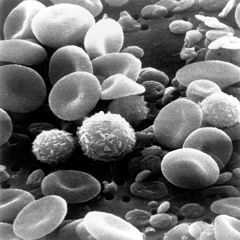
Erythrocytes, or red blood cells, are the most abundant cells in human blood. They carry oxygen from the lungs to the body’s tissues and return carbon dioxide from the tissues back to the lungs. Under the microscope, they appear as biconcave discs without a nucleus. This unique shape increases their surface area for oxygen transport.
Leukocytes (White Blood Cells)
Leukocytes, or white blood cells, are the warriors of the immune system. They defend the body against infection and disease. There are various types of white blood cells, each with different roles. Some attack invaders directly, while others produce antibodies. Under the microscope, they are larger than red blood cells and have a nucleus.
Thrombocytes (Platelets)
Thrombocytes, or platelets, are cell fragments that play a crucial role in blood clotting. If a blood vessel is damaged, platelets gather at the site, form a plug, and prevent excessive bleeding. They are much smaller than red or white blood cells and have a unique, irregular shape.
Plasma
Plasma is the liquid part of blood. It carries blood cells, nutrients, hormones, and waste products throughout the body. It also contains proteins that are essential for clotting and immune responses. Under the microscope, plasma is the clear, straw-colored fluid that suspends the blood cells.
By examining blood cells under microscope, health professionals can assess the health and function of these components. Such examination aids in the diagnosis and management of a wide range of health conditions. Furthermore, it serves as a base for further medical research and advancements in treatment strategies.
Preparing a Blood Sample for Microscopic Examination
Before observing blood cells under microscope, the preparation of the blood sample is critical. This process ensures that the microscopic images are clear and that the cells’ characteristics are discernible. Here’s how lab technicians typically prepare a blood sample for examination:
- Collection: A healthcare professional collects a blood sample from the patient, usually by venipuncture.
- Anticoagulation: To prevent clotting, they mix the blood with an anticoagulant. This step is crucial for preserving the blood’s natural state.
- Smear Preparation: The technician places a drop of blood on a glass slide. They then use another slide to spread the blood across the slide’s surface, creating a ‘blood smear’.
- Staining: After the smear dries, they apply specific stains to the slide. Stains like Wright’s or Giemsa make different blood cells more visible under the microscope.
- Fixing: The slide is then heated or treated with methanol. This process ‘fixes’ the cells to the slide and locks in the stain.
- Observation: Finally, the prepared slide is placed under the microscope. Technicians adjust the focus to view the blood cells in detail.
Each of these steps is crucial for achieving accurate and reliable results when examining blood cells under microscope. Proper sample preparation reveals the intricate details of erythrocytes, leukocytes, and thrombocytes, allowing for a thorough analysis.
Erythrocytes: Observing Red Blood Cells
When we delve into blood cells under microscope, erythrocytes or red blood cells present a spectacle. These cells are vital for our survival. They transport oxygen to body tissues and bring back carbon dioxide to the lungs. This process is essential for sustaining life.
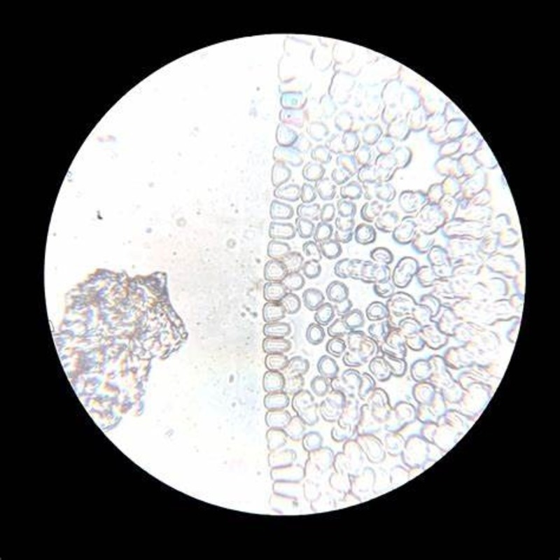
Under the microscope, erythrocytes appear as small, biconcave discs. This unique shape is a marvel of nature. It allows for a greater surface area to volume ratio. This in turn enhances the cells’ ability to ferry oxygen.
In a healthy individual, erythrocytes are numerous and maintain a uniform size and shape. But, under microscope variations can become apparent. These changes may signal anemia or other blood-related disorders.
Pathologists count and study these cells’ characteristics to assess a person’s health. They look for color, size, and the absence of nuclei in the cells. Each detail can point to a different aspect of the patient’s health status.
When preparing a blood sample, care is taken to preserve erythrocytes’ integrity. This ensures accurate observations. Observing red blood cells under microscope is not only critical for diagnosis. It also provides rich information about the body’s physiological state.
Leukocytes: Identifying White Blood Cells
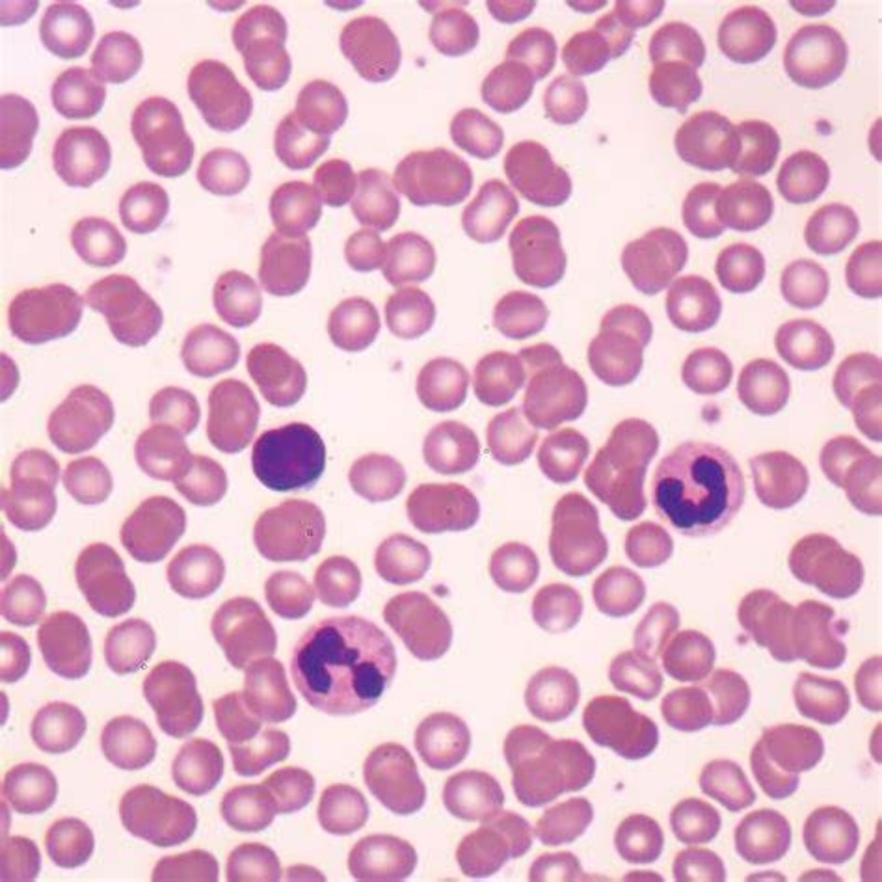
Leukocytes, also known as white blood cells, are the defenders of our immune system. These cells are essential for fighting off infections and other diseases. When looking at blood cells under microscope, leukocytes stand out due to their larger size and the presence of a nucleus, which differentiates them from the erythrocytes.
Under the microscope, one can distinguish several types of leukocytes, each with a unique role in the immune response. These include lymphocytes, which produce antibodies; neutrophils, which target bacteria; eosinophils, which combat parasites; and monocytes, which handle dead or damaged cells.
By observing these cells, pathologists can gather critical information about a patient’s immune system status. For instance, an increased number of leukocytes might be a sign of an infection or an inflammatory condition. Conversely, a lower count can point to possible complications, such as a compromised immune system.
In the staining process of preparing a blood sample, leukocytes are stained so that their components, such as the nucleus, can be easily seen. This is vital for identifying any abnormal shapes, sizes, or other characteristics that could indicate health issues.
The meticulous examination of leukocytes under microscope not only aids in diagnosing illness but also in monitoring the effectiveness of treatments. As a crucial component of the blood, white blood cells under microscope provide a window into the body’s state of immunity. By understanding these cells better, healthcare professionals can enhance patient care significantly.
Thrombocytes: Examining Platelets
When studying blood cells under a microscope, thrombocytes, or platelets, demand special attention. Their role in clotting is vital to prevent bleeding. Platelets often stick together at injury sites, forming clots to stop blood flow.
Under the microscope, platelets appear as small, irregularly shaped cell fragments. Unlike red and white blood cells, they do not have a nucleus. Spotting them requires a keen eye, as they are much smaller. Platelets clump together, which is key in stopping bleeding from wounds.
A normal platelet count is crucial for health. Too many platelets can lead to clots where they’re not needed. This can cause strokes or heart attacks. Too few platelets mean bleeding may not stop as it should. This can be dangerous, especially with cuts or internal injuries.
Pathologists count platelets to check for disorders. Counts that are too high or low can indicate an issue. They also look at their shape and how they cluster together. Variations can show signs of diseases that affect clotting.
By examining platelets carefully, doctors can better understand clotting problems. This helps them guide treatment for conditions like thrombocytopenia or hemophilia. In these conditions, clotting doesn’t work right, leading to health risks. Observing platelets under the microscope is thus key to preventing and treating clotting disorders.
Common Blood Cell Disorders and Their Microscopic Features
The microscopic examination of blood cells can reveal disorders that affect blood health. By looking at blood cells under a microscope, doctors can spot changes. These changes include variations in cell size, shape, and number. Here we explore some common blood cell disorders and what they look like under the microscope:
Anemia: This is a condition where there are not enough red blood cells. It can cause fatigue and weakness. Under the microscope, red blood cells may look pale and could be varied in size. This is known as anisocytosis.
Leukemia: A type of cancer that affects white blood cells. It leads to a high number of abnormal white blood cells. These cells often appear disorganized and can be of different stages of development.
Thrombocytopenia: A disorder where platelet counts are low. It increases bleeding risk. Microscopically, there are fewer platelets seen, and they may vary in size.
Polycythemia Vera: A rare disorder that leads to too many red blood cells. It can cause headaches and dizziness. The cells can look overcrowded and more intense in color.
Infectious Mononucleosis: Often caused by the Epstein-Barr virus, it shows a high number of white blood cells. Some of these cells under the microscope look larger and have atypical shapes.
Each disorder has unique features when viewed through a microscope. Spotting these features helps in diagnosing and guiding treatment. The understanding of these microscopic signs is key for healthcare professionals. With this knowledge, they can manage and treat these conditions more effectively.
Technological Advancements in Blood Cell Imaging
As an SEO expert and professional blogger focusing on the microscopic examination of blood cells, it is crucial to keep abreast of the technological advancements that have revolutionized this field. While traditional microscopy has provided us with detailed views of blood cells under microscope, new techniques and technologies have emerged, enhancing our capabilities to observe and analyze these vital components of our health.
In recent years, advancements in digital imaging and high-throughput screening have made it possible to capture images of blood cells with stunning clarity and detail. Digital microscopes equipped with powerful cameras and image analysis software allow for quicker and more accurate assessments of blood cell morphology. These tools have improved the efficiency of detecting abnormalities in cell size, shape, and population.
In addition to these, automation and artificial intelligence (AI) have also begun to play significant roles. Automated systems are now able to prepare and examine blood samples with minimal human intervention, reducing the potential for error and ensuring consistent results. AI algorithms go a step further by learning from vast sets of data to identify patterns and anomalies that might escape the human eye.
These technological advancements in blood cell imaging are not only improving the accuracy of diagnoses but are also expediting the process, leading to quicker patient care. As we continue to embrace these technologies, our understanding of blood cell behavior and our ability to diagnose and treat blood-related disorders will undoubtedly advance.


