Anatomy of the Skin
Understanding the structure of the skin is foundational in exploring its health under the microscope. The skin is not just a single layer; it’s a complex organ made up of several layers that each play crucial roles in protection, sensation, and regulation.
The Epidermis Layer
The epidermis is the outermost layer of the skin. When looking at skin under a microscope, it’s the first layer we encounter. It’s mainly composed of keratinocytes and is responsible for the skin’s barrier function. The epidermis also contains melanocytes, which contribute to skin color and protect against UV rays. Regular examination of this layer can reveal conditions like psoriasis or eczema.
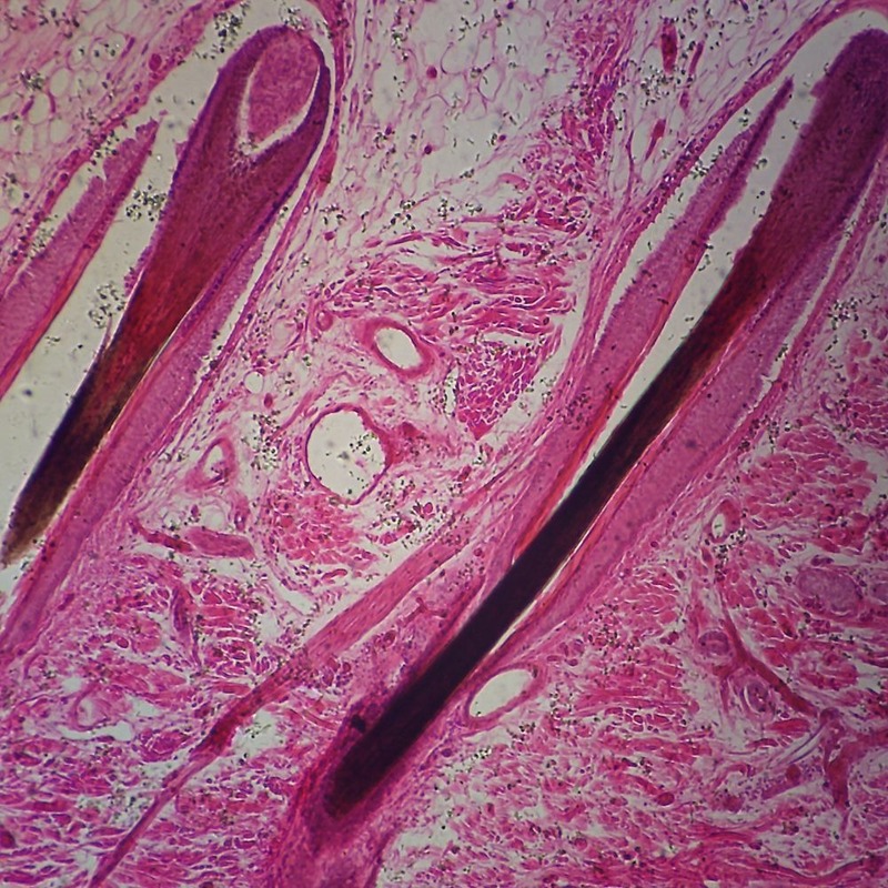
The Dermis Layer
Beneath the epidermis lies the dermis. This complex layer is rich in collagen and elastin, providing the skin with strength and elasticity. Hair follicles, sweat glands, and blood vessels are also found here. The dermis is key to identifying signs of aging and damage when scrutinized under a microscope.
The Hypodermis Layer
The deepest layer is the hypodermis, also known as the subcutaneous tissue. This layer contains fat cells that help insulate the body and act as a shock absorber. Observing this layer under a microscope can aid in understanding conditions related to fat deposits or the assessment of certain treatment effects.
The Role of Microscopy in Skin Examination
Microscopy plays a pivotal role in examining skin health. By magnifying skin under a microscope, professionals can observe details unseen by the naked eye. This tool is vital for dermatologists and researchers. They rely on it to diagnose conditions, study skin’s response to treatments, and understand the aging process.
Types of Microscopes Used
Different microscopes offer various levels of detail. The most common ones are light microscopes, which light up skin samples to show the basic structure. Dermatologists often use these for a quick assessment. But for deeper insights, electron microscopes come into play. They show skin at a cellular level, revealing much finer details. For dynamic analysis, confocal microscopes create 3D images of living cells in the skin. Each type of microscope serves a specific function in skin analysis.
Preparing Skin Samples for Microscopy
Before examining skin under a microscope, proper preparation is key. This involves cleaning the skin area to remove oils and debris. After cleaning, the skin is usually biopsied with a small tool. The sample is then fixed with chemicals to preserve the tissue. It’s often stained to highlight different components. Once prepped, the sample is placed on a slide for viewing. Careful preparation ensures accurate and useful results from the microscopic examination.
Identifying Common Skin Conditions
When viewing skin under a microscope, various common skin conditions become identifiable. By examining skin cells and tissue structures, professionals can diagnose and understand the nature of these conditions.
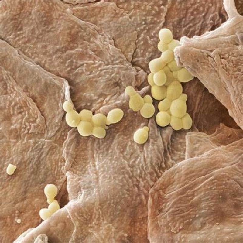
Bacterial Infections
Bacterial infections in the skin appear as clusters of abnormal cells. These can sometimes be accompanied by pus or other signs of infection. Under the microscope, the presence of bacteria can be confirmed, providing clear evidence for diagnosis.
Fungal Infections
Fungal infections are characterized by the presence of fungal hyphae or spores. These elements have distinctive shapes and sizes, making them visible when skin is magnified. They are often found within or beneath the epidermal layer.
Viral Infections
Viral infections often alter the appearance of skin cells, such as causing them to balloon or cluster together. These changes provide visual clues to the presence of a virus within skin tissue when viewed under a microscope.
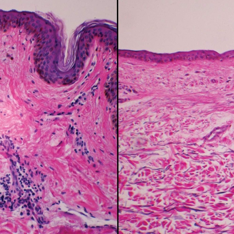
Inflammatory Skin Diseases
Inflammatory skin diseases, such as psoriasis and eczema, show increased cell activity under a microscope. Signs include thickened epidermis and infiltration of immune cells, which suggest an ongoing inflammatory response.
Analyzing Skin Aging at the Microscopic Level
As skin ages, changes occur at a cellular level. These changes are not visible to the naked eye. However, using a microscope can bring these subtle shifts into sharp focus.
Cellular Changes with Age
When we examine skin under microscope, we can see cells gradually lose their robust nature. For instance, fibroblasts that produce collagen slow down. This leads to less firm skin. Skin cells also turn over more slowly which causes a buildup of older cells on the surface. These changes can signal the onset of wrinkles and a loss of youthful appearance.
The Impact of UV Radiation
UV radiation is notorious for accelerating skin aging. Microscopic examination reveals UV light can damage the DNA in skin cells. This damage can prevent cells from functioning well. Over time, it contributes to aging signs like sunspots and loss of skin elasticity.
The Role of Collagen and Elastin
Collagen and elastin are vital for skin’s firmness and stretch. As we age, their production declines, visible under a microscope as thinner, less dense fibers. This change is a major reason why skin sags and wrinkles form. It’s essential to monitor the condition of these proteins to understand skin aging comprehensively.
The Skin’s Response to Treatments Under the Microscope
When exploring how skin responds to treatments, a microscope can offer invaluable insights. It shows us what happens at the cellular level when skin receives various therapeutic interventions. Here’s how some common treatments appear under magnification:
Topical Medications
Microscopic observation of skin treated with topical medications reveals a lot. These meds can change how cells look and function. For instance, anti-inflammatory creams reduce redness and swelling. They do this by affecting the cells involved in inflammation. Similarly, we see hydrating agents fill the gaps between cells. This makes the skin look plump and smooth.
Laser Therapy
Lasers do more than just target blemishes. Under the microscope, we see lasers stimulate collagen production. The heat from the laser helps to tighten the skin structure. It also removes damaged skin cells. This process leaves behind a fresher and more youthful layer.
Chemical Peels
Chemical peels are another treatment visible under a microscope. They remove the top layer of the skin. This reveals a new layer underneath that’s smoother and less damaged. Microscopy shows how peels can reduce fine lines and sun damage. They work by stripping away older cells that hold signs of aging. This encourages new cell growth. It’s a powerful way to rejuvenate the skin’s appearance.
Skin Hydration and Microscopy
Hydration is a key factor in skin health. Under a microscope, we can spot signs of well-hydrated skin versus dehydrated skin. This knowledge helps us understand and address hydration issues.
Understanding the Microscopic Signs of Dehydration
When we inspect skin under microscope for dehydration, we see several tell-tale signs. Dull and less supple skin cells are common. You might also see gaps between skin cells. These gaps should be minimal in well-hydrated skin. Hydrated skin cells are plump and tightly packed together. They form a strong barrier against environmental stressors. On the flip side, dehydrated skin cells can contribute to irritation and sensitivity. They make the skin look flaky and feel rough to the touch. Recognizing these patterns is crucial. It allows for timely treatment with moisturizers and hydrating serums.
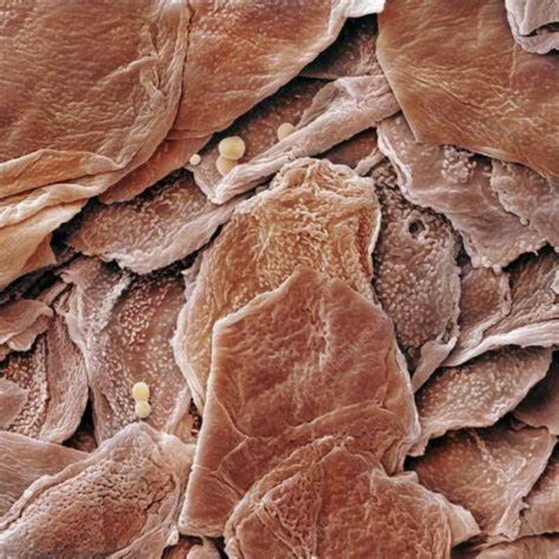
The Role of the Stratum Corneum
The stratum corneum lies at the top of the epidermis. It plays a huge part in skin’s hydration. This layer holds lipids and natural moisturizing factors. These elements prevent water loss and protect against harmful microbes. Under a microscope, a healthy stratum corneum appears even and intact. In contrast, dehydration can result in a disrupted barrier. We might see cracks or excessive shedding of skin cells. Restoring the integrity of the stratum corneum is vital for proper hydration. It helps the skin to retain moisture and stay healthy.
Innovations in Skin Microscopy
The field of skin microscopy is constantly evolving. Recent advancements have pushed the boundaries of what we can see when examining skin under a microscope. These innovations have crucial roles in dermatology and cosmetic science. They offer clearer understandings of skin health, aging, and disease. Let’s look closer at some cutting-edge microscopy techniques used today.
Confocal Microscopy
Confocal microscopy stands out among skin imaging technologies. It lets us see live tissue at microscopic levels without the need to cut and prepare samples. This method uses a laser to scan skin and create high-resolution images. It reveals the structure and function of skin cells in real-time. Dermatologists use it to diagnose diseases and watch how skin reacts to treatments on the spot.
Multiphoton Microscopy
Multiphoton microscopy brings us even deeper insights. It emits bursts of infrared light that penetrate skin layers without damaging them. This technique captures images of deeper skin structures. It shows us how cells interact in their natural environment. It is helpful for looking at collagen and elastin in the dermis. Researchers use it to study the aging process and the effects of treatments over time.
Optical Coherence Tomography
Optical coherence tomography (OCT) is like an ultrasound for the skin. It uses light waves to take cross-sectional pictures of the skin. OCT can map out layers of skin and spot changes in tissue structure. It is useful for detecting skin cancers and other conditions. This non-invasive method provides a quick look at skin health. It does so without the need for biopsies or staining samples.
These innovations in microscopy enhance our ability to study skin under microscope. They provide dynamic, detailed images of skin, from its surface down to the cellular level. As technology progresses, we can expect even more precise tools for exploring skin health.


