Introduction to Cell Microscopy
The study of cells under microscope has revolutionized our understanding of biology. In a glimpse, it reveals an intricate world, invisible to the naked eye. From observing the basic structure to identifying complex processes, cell microscopy offers profound insights into life’s building blocks.Discover how to view cell under microscope! Learn about preparation, techniques, and the differences between plant and animal cells.
Microscopy has empowered scientists to view details within cells. It allows for the differentiation of cell parts, such as the membrane, nucleus, and organelles. This visualization is crucial for studying cellular functions and diseases.
Through the use of various microscopy techniques, researchers can not only observe cells in detail but also track their behaviors over time. Students and professionals worldwide utilize cell microscopy to expand their knowledge. It is essential in fields such as genetics, pharmacology, and microbiology.
For anyone embarking on this microscopic voyage, understanding the tools and methods is key. This journey will take us from the foundational light microscopes to the advanced electron and fluorescence microscopes. As we delve deeper, we’ll uncover the roles of each type in revealing the secrets of cells.
Whether you are a student, researcher, or simply curious, the world of cell microscopy beckons. Its revelations are both fascinating and invaluable to science. Together, let’s explore this hidden universe and its profound impact on our comprehension of life.
The History of Microscopy and Cell Observation
Tracing back to the 17th century, the history of microscopy begins with the invention of the first compound microscope. This groundbreaking device allowed scientists like Robert Hooke to view cells for the first time. Hooke’s work led to the coining of the term ‘cell’ after observing the cell walls of a cork.
The development of cell observation evolved rapidly. Antonie van Leeuwenhoek, using his handcrafted microscopes, discovered bacteria, free-living and parasitic microscopic protists, sperm cells, blood cells, and much more. His discoveries laid the groundwork for microbiology.
Over centuries, microscopes have become more sophisticated. The 19th century brought refinements in lens quality and the introduction of the achromatic lens, reducing distortion and improving image clarity. It was during this era that the theory of cells as the basic unit of life took form.
The 20th century unlocked a new world with the electron microscope. This microscope used a beam of electrons, rather than light, to create images. It magnified structures up to two million times, unveiling details unseen with traditional light microscopy.
As microscopy techniques advanced, so did our knowledge of cells and their functions. The discovery of organelles, enzymes, and the complex interactions within cells has been made possible by continuous improvements in microscopy.
To this day, scientists rely on the legacy of these early pioneers and their instruments to explore life at the cellular level. Cell under microscope studies remain a centerpiece in biological research, crossing over into numerous scientific disciplines.
Types of Microscopes Used for Viewing Cells
When delving into the cellular world, various types of microscopes come into play. Each type has its own unique features and applications, enabling scientists to observe cells in different ways. Here is a closer look at the main types of microscopes used in cell observation.
Light Microscopy
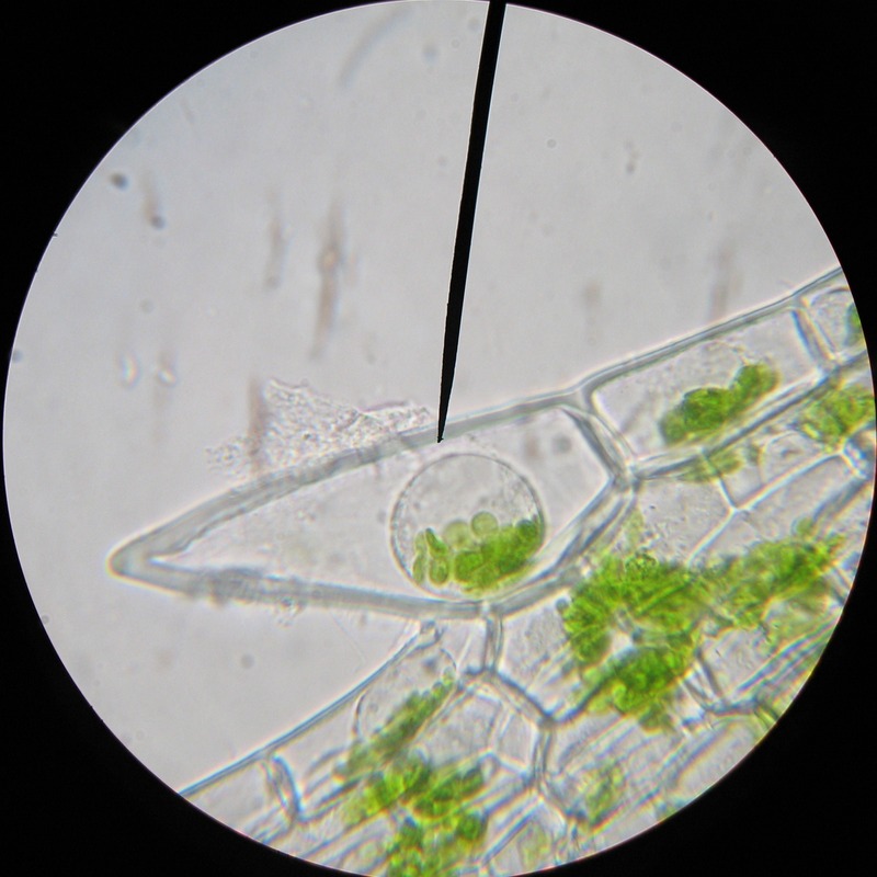
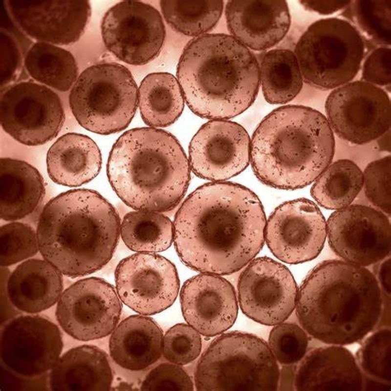
Light microscopy involves using visible light to illuminate specimens. It’s the oldest and most commonly used method to view cells. Often referred to as optical microscopy, it encompasses several techniques:
- Brightfield microscopy, where light passes directly through the sample, making the cell components visible against a bright background.
- Darkfield microscopy, which enhances contrast in unstained cells, highlighting them against a dark background.
- Phase-contrast and differential interference contrast (DIC) microscopy, which improve the visibility of transparent specimens by enhancing differences in refractive index.
These methods are ideal for live cell observation, as they usually don’t harm the specimens.
Electron Microscopy
Electron microscopy uses beams of electrons instead of light to create an image. It includes two main types:
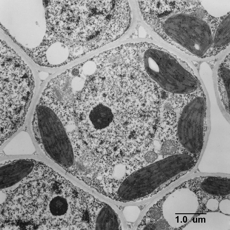
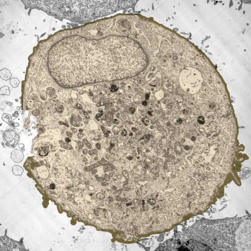
- Transmission electron microscopy (TEM), which provides detailed images of the inner structure of cells. It allows researchers to see organelles within the cytoplasm.
- Scanning electron microscopy (SEM), which offers a three-dimensional view of cell surfaces.
This type of microscopy can achieve higher magnifications and better resolution than light microscopes, revealing the smallest details of cell structure.
Fluorescence Microscopy
Fluorescence microscopy harnesses the power of fluorescent molecules to illuminate specific parts of the cell. Cells or their components are stained with fluorescent ‘tags’ that emit light when excited by a specific wavelength. This technique enables the observation of dynamic processes in cells and localization of proteins and other molecules. It’s particularly useful in molecular biology and genetic research.
While these types of microscopes are most prevalent, there’s a vast array of specialized microscopic technologies that scientists can employ to explore cells under microscope. Each plays a crucial role in progressing our understanding of the cellular universe.
Preparing Cells for Microscopic Observation
Before a cell sketch comes to life under a microscope, proper preparation is crucial. This process ensures that observations reflect the cell’s true structure and function. There are several steps involved in preparing cells for microscopic observation.
First, cells often need to be isolated from a sample. This might mean detaching cells from a tissue or filtering out unwanted particles. Isolation helps to get a clear view of the cell itself.
Second, fixation is key. This step preserves cell structure by stopping all biological processes. Chemicals like formaldehyde are common fixatives. They lock everything in place, allowing the sample to withstand further processing steps.
Third, staining is applied to many cell preparations. Dyes like Hematoxylin and Eosin (H&E) give contrast to structures within the cell. Stains can highlight specific elements, such as DNA, proteins, or lipids.
Mounting is the next step, where the cell is placed on a slide and covered with a cover slip. This protects the cell and helps keep it in focus during observation.
Sample thickness also matters. Slicing techniques like sectioning can cut a sample into thin layers. Thin samples let light or electrons pass through, revealing detailed cell structures.
Lastly, the choice of medium in which the sample will be placed can affect visibility. Some preparations require a certain environment to maintain cell integrity and enhance the image quality.
Each of these steps needs to be carried out with precision. Any error might distort the image or misrepresent the cell’s components. Thus, scientists take great care during preparation to ensure accurate and useful observations under the microscope.
Cell Structure and Organelles Revealed Under the Microscope
Once cells are prepared and placed under the microscope, their complex structure becomes revealed. This view opens up a detailed perspective on how life functions at a cellular level. Let us explore the various components that become visible when we observe a cell under microscope.
Visible parts of the cell include the cell membrane, which serves as a protective barrier. It controls what enters and leaves the cell. Inside, the nucleus stands out as the control center, housing DNA, the blueprint of life. The cytoplasm, filled with a jelly-like fluid, holds various organelles each with a special role.
Mitochondria, known as the powerhouses, provide the cell with energy. The Endoplasmic Reticulum (ER) comes in two forms, rough and smooth, and plays a part in making proteins and lipids. Ribosomes, small dots scattered in the cell, read genetic instructions to create proteins. The Golgi apparatus modifies and packs proteins for transport.
Other organelles include lysosomes that break down waste and vacuoles that store substances. Each organelle is crucial for cell survival and function. Under the microscope, even tiny structures get magnified, showing the intricate and efficient organization of life at the cellular level.
In addition to these standard components, special cells may have unique parts. For example, plant cells contain chloroplasts which perform photosynthesis. Similarly, nerve cells have long extensions to transmit signals. By studying cells under the microscope, we continue to discover new aspects of their structure and role.
Microscopy not only allows us to see the parts of a cell but also to watch these components work together. It shows us the dynamic processes that sustain life. As we observe cells, we gain a deeper appreciation for the complexity and beauty of the biological systems that make up living organisms.
Application of Cell Microscopy in Research and Medicine
The use of cell under microscope has vast applications in both research and medicine. In research, it enables scientists to unravel the complex mechanisms within living organisms. For instance, cell microscopy aids in the study of cellular processes such as mitosis and meiosis. It’s essential in genetics research, helping to identify how traits are inherited and how genetic disorders occur.
In medical fields, this technology is pivotal for diagnosing diseases. Pathologists often rely on cell microscopy to examine tissue samples. This examination helps them to detect cancerous cells, identify infections, and understand inflammatory diseases. Microscopy is also used in developing new treatments. Researchers can observe how drugs affect cells – noting changes in cell structure, growth, or death. This kind of observation is key in pharmacology, particularly in drug discovery and development.
Moreover, in the realm of microbiology, microscopy allows for the observation of microscopic organisms. This insight is crucial for understanding infectious diseases and devising ways to combat them. By observing cells under microscope, scientists can see how bacteria and viruses invade cells or how the immune system responds to these threats.
In the field of regenerative medicine, cell microscopy is instrumental. It’s used to monitor stem cell differentiation and to ensure the quality of tissues grown in the lab. This close monitoring is essential for developing therapies that one day may regenerate damaged organs or tissues.
Overall, cell microscopy is a cornerstone of contemporary scientific research and medical practice. It has transformed our understanding of life at the cellular level and continues to be a window into the hidden universe within us.
Advances in Microscopic Techniques and Future Outlook
The field of microscopy is in a constant state of evolution. As technology progresses, new microscopic techniques emerge, offering even deeper insights into the cell’s intricacies. These advances not only enhance image quality but even allow real-time observation of cellular processes.
One significant advancement is the development of super-resolution microscopy. This technique surpasses the diffraction limit of light, revealing details smaller than previously possible. Techniques such as STED (Stimulated Emission Depletion) and PALM (Photo-Activated Localization Microscopy) provide images with nanometer resolution.
Another cutting-edge approach is live-cell imaging. Here, scientists watch cells in action, observing processes like division and migration unfold over time. This method requires gentle imaging conditions to keep cells alive and healthy during examination.
3D imaging technologies have also made great strides. They reconstruct detailed three-dimensional models of cells, giving us a more complete view of cell architecture. Research in tissue engineering and drug development benefits greatly from this advancement.
Artificial intelligence (AI) is shaping the future of microscopy as well. AI algorithms quickly analyze vast amounts of image data, detect patterns, and even predict cellular behavior. This innovation accelerates research and can lead to breakthroughs in disease diagnosis.
Looking ahead, the adoption of even more precise and sophisticated imaging methods is expected. These methods will refine our ability to see cells at the molecular level. As we advance, we hope to gain a more comprehensive understanding of cellular function and disease.
In conclusion, with every microscopic technique improvement, we step closer to unlocking the deepest secrets of the cell under microscope. This progress holds promising potential for scientific discoveries that can change our view of life and medicine.
Conclusion: The Impact of Cell Microscopy on Science
The study of cell under microscope has deeply changed science. It allows us to see the unseen, unravelling the mysteries of the smallest life units. This journey through microscopes has been crucial for countless discoveries across all biology fields.
Microscopy has shaped our knowledge about life itself. It helps us understand how cells function, and how they form the bases of complex organisms. From learning about organelles to watching cells divide, microscopy offers us a front-row seat to the action.
In research, cell microscopy is a key tool. It helps us study diseases, develop medicines, and much more. The findings from this small world have a big impact. They guide scientists in many areas like genetics, pharmacology, and microbiology.
In medicine, cell microscopy helps diagnose and treat illnesses. By studying cells, doctors can find cancer early and track infections. Drug testing under the microscope leads to better and safer medicines.
As we continue to develop new microscopy methods, we can hope for more. Better images and real-time cell watching could lead to huge advances. This progress can change our understanding of health and disease.
In summary, the power of viewing a cell under microscope shapes modern science. It will keep offering insights for future researchers and medical experts. This tiny window into the unseen world gives us a vast universe of knowledge to explore.


