What Is Cardiac Muscle
Cardiac muscle is a specialized type of muscle found only in the heart. Its unique structure allows it to beat and pump blood throughout the body without tiring quickly. Unlike skeletal muscle, which we can control, cardiac muscle works involuntarily. This means it does its job without us having to think about it. Its cells, called cardiomyocytes, join to form a strong, yet flexible network. This network stretches and contracts to make the heartbeat. Its durability and endurance are remarkable. With each beat, blood gets pushed to our organs and tissues, keeping us alive. A look at cardiac muscle under microscope reveals its complexity and beauty. We’ll explore its traits and roles in more detail as we move through the blog.
Characteristics of Cardiac Muscle Cells
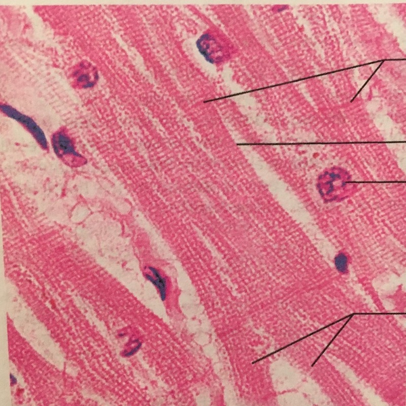
Cardiac muscle cells, or cardiomyocytes, show distinctive features, especially when viewed under a microscope. Each cell has a single central nucleus. This makes them easily distinguishable from other muscle cell types. The cells contain rich, densely packed mitochondria. These powerhouses supply the energy needed for the heart’s constant activity. Furthermore, cardiac cells have a branched structure. It enables them to connect with each other closely. This unique formation forms a tight network. The network helps in the synchronized contraction of the heart.
Another remarkable trait is the presence of intercalated disks. These structures bind cardiomyocytes together and support electrical conductivity. This allows for swift communication between cells. Thus, the cells work in harmony, making the heartbeat as one unit. Cardiac muscles also possess a high amount of myoglobin. The myoglobin stores oxygen, which is critical for maintaining the heart’s stamina.
Through advanced staining techniques and powerful microscopes, these intricate features of cardiac muscle cells become visible. They underline the sophisticated nature of the heart’s design. The resilience and efficiency of these cells ensure the heart beats tirelessly, sustaining life.
Cardiac Muscle Structure and Function
Cardiac muscle structure showcases a complex yet efficient design. Its function is crucial for life. Cardiac muscles are made of individual cells known as cardiomyocytes. These cells connect and form a network. The network allows for synchronized heartbeats. Every contraction pushes blood through the body.
The heart’s structure aids its relentless task. The walls of the heart have three layers. The inner layer, or endocardium, comes into contact with the blood. The middle layer, the myocardium, contains the cardiac muscle. This is where the power of contraction originates.
The outer layer, the epicardium, protects and provides lubrication. The function of cardiac muscles goes beyond contraction. It helps maintain blood pressure and ensures timely blood flow. Each muscle’s rhythmic contraction is a life-sustaining dance.
The heart’s valves manage blood direction. They make sure blood flows one way and prevents backflow. Efficiency is key in cardiac muscle function. Tiny capillaries deliver oxygen to the continuously working muscles.
Electrical impulses guide the timing of each heartbeat. These impulses pass through the muscle network with precision. This coordination ensures that the heart functions as one unit. A strong, coordinated heartbeat is vital. It is what allows the rest of the body to function properly.
Cardiac muscle under microscope reveals how all these functions are rooted in intricate cell design. Even at this microscopic level, the complexity and precision of the cardiac muscle are evident.
The Role of Cardiomyocytes in the Heart
Cardiomyocytes, often referred to as heart muscle cells, play a pivotal role in cardiovascular health. Their primary duty lies in contracting and relaxing, which facilitates the heartbeat. Each cardiomyocyte works in unison with surrounding cells to form a powerful, rhythmic force. This force pumps blood throughout the entire body. These cells are quite remarkable, as they combine strength, endurance, and precision.
By interacting through intercalated disks, cardiomyocytes synchronize their contractions. This synchronization is crucial for a steady, strong heartbeat. They ensure that each heartbeat is efficient and consistent. Moreover, cardiomyocytes are self-excitable. This means they can generate their own electrical impulses. These impulses trigger their contractions and regulate the heart’s rhythm.
Cardiomyocytes also adapt to the body’s changing needs. For instance, during exercise, they contract more often to increase blood flow. When at rest, they slow down, conserving energy. This adaptability is essential for maintaining homeostasis.
In health conditions affecting the heart, cardiomyocytes can get damaged. This damage impacts their ability to contract effectively. Studying cardiac muscle under microscope helps researchers understand these conditions better. It allows them to observe changes in cardiomyocytes at a cellular level. This microscopic insight is valuable for developing treatments for heart diseases.
Without cardiomyocytes and their synchronized work, the heart would fail to function. Thus, their role is irreplaceable in keeping the heart beating and the body alive.
Visualizing Cardiac Muscles: Tools and Techniques
To thoroughly study cardiac muscle under microscope, scientists employ a variety of tools and techniques. The main aim is to reveal the intricate details of these muscles at the cellular level. Here are some of the key methods:
- Light Microscopy: This traditional approach uses visible light to magnify cardiac muscle tissue samples.
- Electron Microscopy: For greater detail, electron microscopy offers higher magnification and resolution than light microscopy. It’s ideal for observing the complex structure of cardiomyocytes.
- Fluorescence Microscopy: This technique uses fluorescent stains to highlight specific parts of cells, such as proteins or organelles. It is particularly useful for studying live cell processes.
- Confocal Microscopy: Confocal microscopy creates sharp images by focusing a laser on thin slices of tissue. It helps observe the structure of cardiomyocytes in three dimensions.
- Immunohistochemistry: This method detects specific proteins within cardiac muscle cells. Antibodies bind to these proteins and are then visualized using dyes.
- Atomic Force Microscopy: To measure the mechanical properties of cells, atomic force microscopy applies a very small force to the cell surface.
Each of these tools plays a vital role in visualizing the function and form of cardiac muscles. Techniques like staining and tagging allow scientists to track changes and activities within living cells. By leveraging these techniques, researchers can gain crucial insights into how cardiac muscles work and how they are affected by various diseases. Understanding cardiac muscle at the microscopic level can lead to better diagnostics and treatments for heart conditions.
Observing Cardiac Muscle Activity Through Microscopy
Observing cardiac muscle activity under a microscope unfolds a world of dynamic processes. In the lab, scientists closely monitor how these muscles function. They use high-powered microscopes to watch the heart cells work. This deep look into the cardiac muscle reveals the rhythm of life in each beat.
To capture cardiac muscle activity, advanced imaging techniques are essential. Here’s how they do it:
- Live Cell Imaging: This lets researchers see cardiomyocytes in action. They observe how cells contract and relax over time.
- Time-lapse Photography: By taking pictures at intervals, a video of the heartbeat cycle is made. This shows how cardiac muscles coordinate their contractions.
- Phase-contrast Microscopy: This method helps visualize cell movement without dyes. It makes the beating heart cells stand out against their background.
- Electrophysiological Recording: Here, the electric activity that drives the heartbeat is measured. It shows how signals move through cardiac muscles to keep the beat steady.
Each heartbeat under a microscope is a sight to behold. The contraction of the heart is a smooth, continual motion. It begins as electrical signals prompt the cardiomyocytes to compress. Then, they release, allowing the heart to fill with blood again. On the microscopic scale, this pattern is both precise and magnificent.
Scientists gain essential insights by observing these heartbeat mechanisms. They see how different factors, like drugs or diseases, affect the heart. This is key in understanding heart health and guiding treatment options. Watching cardiac muscles work under the microscope underscores their vital role. It shows how they keep the blood coursing through our veins. This knowledge is critical for medical breakthroughs that save lives.
Comparing Skeletal, Smooth, and Cardiac Muscle Under the Microscope
Cardiac muscle under microscope offers a distinct view compared to skeletal and smooth muscles. Each muscle type plays different roles in our bodies and has unique characteristics on a cellular level.
- Skeletal Muscle: These muscles attach to bones and control voluntary movement. Under a microscope, skeletal muscle cells appear as long, cylindrical fibers. They have multiple nuclei located on their periphery. The cells show striations, visible lines of actin and myosin filaments.
- Smooth Muscle: Found in the walls of organs like the stomach or blood vessels, smooth muscle cells are shorter and spindle-shaped. They function involuntarily, meaning they work without conscious control. Smooth muscle lacks the striated appearance found in skeletal and cardiac muscle, contributing to its name.
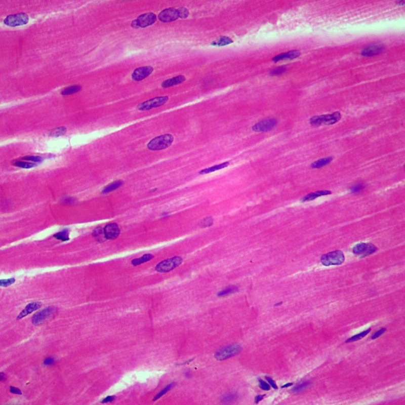
- Cardiac Muscle: Cardiac muscle cells, or cardiomyocytes, are unique to the heart. They feature a single nucleus, are branched, and have intercalated disks for electrical connectivity. The cells contain striations similar to skeletal muscle. But, their arrangement and role are dedicated to heart contractions.
Viewing these muscle types under the microscope shows their distinct structures that reflect their specific functions. Skeletal muscles are built for controlled movement, smooth muscles for involuntary function, and cardiac muscles for a continuous heartbeat. Each type, with its particular design and role, is essential for our survival. By comparing them, researchers can gain a better understanding of how our body’s muscular system operates.
Applications of Studying Cardiac Muscles at the Microscopic Level
Exploring cardiac muscle under microscope has significant applications in medical science. Here are some key benefits:
- Disease Diagnosis: Understanding the microscopic structure of cardiac muscle helps detect heart diseases. Doctors look for changes in cells that signal conditions like cardiomyopathy.
- Drug Testing and Development: Researchers test the effects of new drugs on cardiomyocytes. This helps them see how treatments might work in the human body.
- Gene Therapy Research: Microscopic studies can show how genetic alterations affect the heart. This is key to advancing gene therapy for inherited heart conditions.
- Medical Training: Observing cardiac muscle under a microscope is vital for student doctors. It helps them learn about heart health and disease.
- Improving Surgical Techniques: Surgeons use these insights to refine heart surgeries. They work to ensure minimal damage to heart muscle during operations.
- Public Health Education: Sharing microscopic images can raise awareness about heart health. It can also help teach the importance of lifestyle choices on cardiovascular well-being.
Through studying cardiac muscles at such a tiny scale, scientists can make large strides in health care. This knowledge leads to more accurate diagnoses, better treatments, and improved patient outcomes. The heart is complex, but by delving into its smallest parts, we can protect this vital organ.


