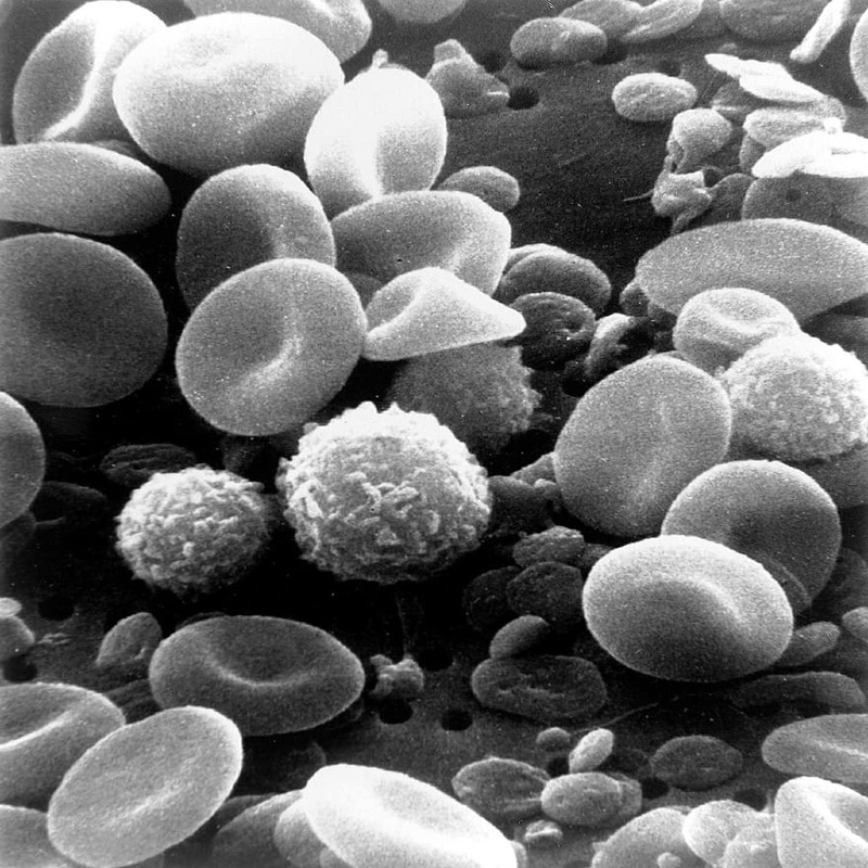
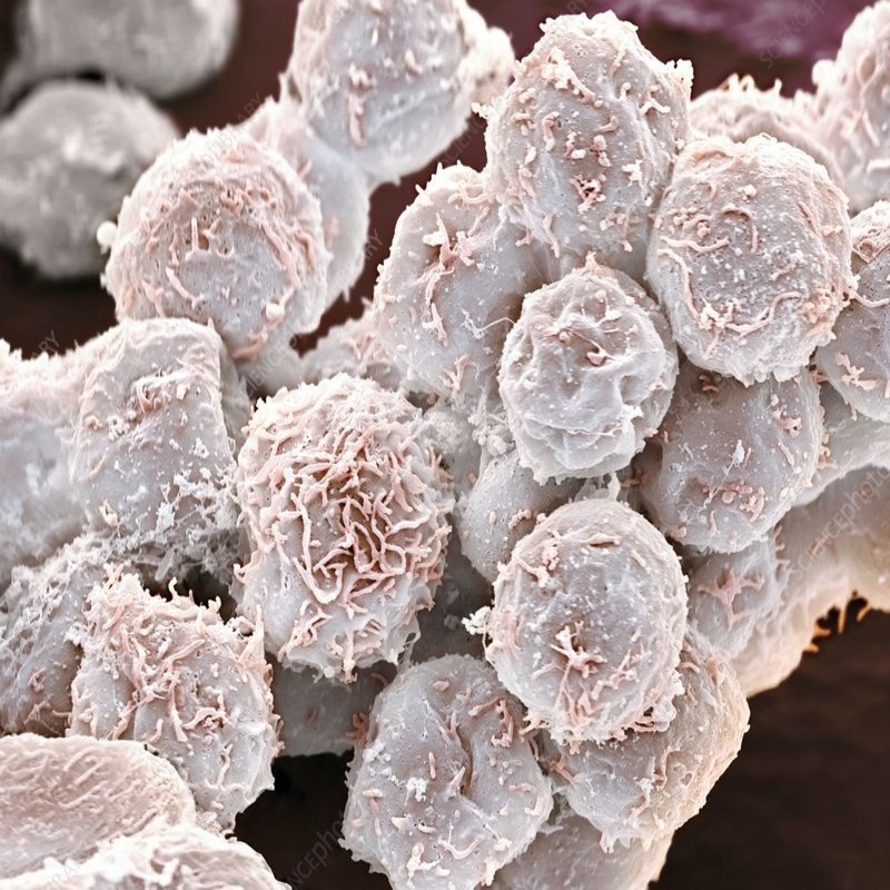
Introduction to White Blood Cells
White blood cells (WBCs) are key players in our immune system. They help fight infections and other diseases. A healthy body has billions of WBCs patrolling the bloodstream. These cells are like small warriors, each specialized in tackling different threats. Viewing white blood cells under a microscope reveals much about our health. By studying WBCs, doctors can diagnose infections, immune disorders, and blood diseases.
Microscopic techniques have evolved to enable detailed WBC examination. Brightfield and phase-contrast microscopy offer basic views. Fluorescence and electron microscopy provide more depth and detail. But first, preparing a blood sample is critical. Well-prepared slides can make a significant difference in visualization quality.
Identifying each type of WBC is also crucial. Our body has several WBC types, each with a distinctive role. Their shapes, sizes, and staining properties vary widely. Learning how different microscopy techniques can highlight these features is essential. Additionally, recognizing when WBCs don’t look normal is important for diagnosis. This introduction will guide you through the intricacies of observing white blood cells under the microscope.
Types of White Blood Cells
Understanding the types of white blood cells under microscope is vital for accurate diagnosis and medical research. Human blood contains five main types of WBCs, each unique in function and appearance.
Neutrophils
Neutrophils are the most abundant type and are first to respond to infections. They destroy bacteria and fungi.
Lymphocytes
Lymphocytes are critical for the body’s immune memory. They have two main types: B cells produce antibodies, and T cells destroy infected cells.
Monocytes
Monocytes are the largest WBCs, engulfing and breaking down pathogens and dead cells. They turn into macrophages in tissues, key for removing debris.
Eosinophils
Eosinophils combat parasitic infections and play a role in allergic reactions. They also help control inflammation.
Basophils
Basophils are the least common but have important roles. They release histamine in allergic responses and help prevent blood clotting.
Each type of WBC can be distinctly identified when viewing white blood cells under microscope, using various staining and microscopy techniques. This differentiation is crucial for doctors to determine the cause of an illness and the best treatment plan.
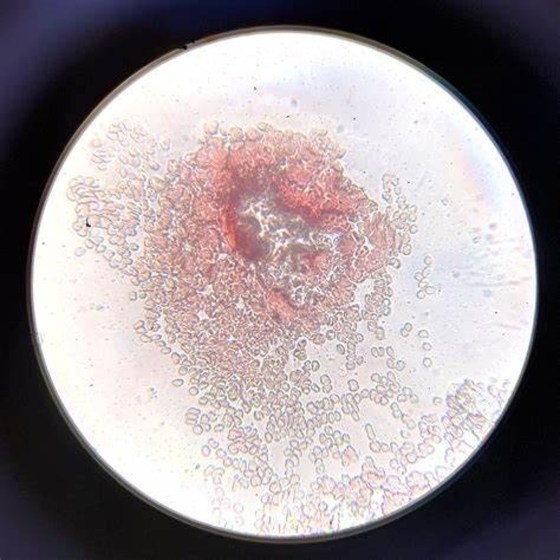
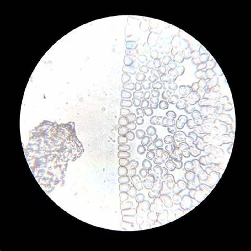
Preparing a Sample for Microscopy
Before viewing white blood cells under a microscope, proper sample preparation is crucial. This process ensures clear and accurate visualization of WBCs. Here’s how to prepare a blood sample for microscopic examination:
- Collection: Draw a blood sample using sterile techniques to prevent contamination.
- Anticoagulation: Mix the blood with an anticoagulant, which stops it from clotting and allows for even spreading on a slide.
- Smearing: Place a drop of blood on a clean microscope slide. Using a spreader slide, create a thin, even blood smear.
- Air-drying: Allow the smear to air dry completely to fix the cells in place. This step is essential for accurate staining.
- Fixation: Fix the sample by using methanol. This preserves the WBCs’ structure during the staining process.
Each step is important to obtain a clear view of the white blood cells under the microscope. Skipping or poorly executing any step can lead to inaccurate results or make it difficult to identify specific types of WBCs. Good preparation contributes to a successful analysis of blood samples and is the first critical step in studying white blood cells using various microscopic techniques.
Microscopy Techniques for Viewing White Blood Cells
After preparing a blood sample, various microscopy techniques can bring the hidden world of white blood cells under microscope into sharp relief.
Brightfield Microscopy
Brightfield microscopy is a basic but widely used technique. Easy to set up, it uses light to illuminate the sample, helping to view unstained or simple stained WBCs. The light passes through the sample, displaying cells against a bright background. While it’s great for general observation, details can be less visible.
Phase-Contrast Microscopy
Phase-contrast microscopy offers more detail. It highlights the contrast between WBCs and their surroundings without staining. This method bends light passing through different parts of the cells, showing their structures clearly. It’s ideal for living cells study, giving insights into WBC behavior.
Fluorescence Microscopy
Fluorescence microscopy uses high-intensity light to excite fluorescent stains in samples. WBCs tagged with these stains emit light, showing up against a dark background. This technique reveals detailed cellular structures and is useful for identifying specific WBC types.
Electron Microscopy
Electron microscopy provides the highest detail level, using electron beams instead of light. Here, electrons interact with the sample, creating extremely detailed images. Although complicated and costly, it shows the finest structures within WBCs, giving unparalleled views of their inner workings. This technique requires careful sample preparation and is used for in-depth cellular research.
Staining Methods for White Blood Cells
To get a detailed look at white blood cells under microscope, staining is key. It gives contrast and helps distinguish between different cell types. Here are common stains used in microscopy:
Wright’s Stain
Wright’s Stain is a mix of eosin and methylene blue. It is perfect for blood smears. This stain dyes WBC nuclei a deep blue-purple and cytoplasm in lighter shades. It helps to see WBCs clearly. Hospitals and labs use it often for its quick and clear results.
Giemsa Stain
Giemsa stain works well for genetic studies and malaria diagnosis. It stains DNA blue. This makes it easier to spot cell types and any changes in chromosomes. The stain also turns the cytoplasm pink or blue, based on the type of WBC. This way, details stand out, aiding in accurate identification.
Immunofluorescence Staining
Immunofluorescence uses antibodies linked to fluorescent dyes. When applied to WBCs, it lights up specific cell components. This type of staining is vital for diagnosing diseases. It can show if WBCs react to different markers. This stain is used for detailed studies and needs special equipment. It gives very specific information about WBC function and structure.
These staining methods are crucial for studying WBCs. They help doctors find and treat diseases with more precision. Each stain has its purpose and ideal use, making white blood cells under microscope easier to analyze.
Identifying Abnormalities in White Blood Cells
Identifying abnormalities while viewing white blood cells under microscope is a critical skill for medical professionals. It involves close examination of the size, shape, and staining pattern of WBCs. Deviations from the norm can signal various health issues. For example, unusually large white blood cells might indicate an infection or leukemia. Atypical staining patterns may suggest genetic conditions or bone marrow problems.
Recognizing Infection Signs
Infections often cause a rise in specific WBC types. Neutrophils, for instance, typically multiply when there’s a bacterial infection. A high eosinophil count can point to a parasitic infection or allergy. By noticing these changes, doctors can diagnose and address the underlying condition sooner.
Diagnosing Blood Disorders
Blood disorders, such as anemia or clotting issues, alter WBC appearance. Pale or oddly shaped leukocytes often raise flags for further testing. Chronic diseases like leukemia are marked by abnormal WBC proliferation. Early detection of these conditions through microscopy is vital to effective treatment.
Detecting Immune Response Problems
Sometimes, WBCs appear normal but don’t function properly. Such cases may indicate immune system disorders. Doctors look for signs like poor WBC response to infections. This requires combining observations with other diagnostic tests to ensure accurate diagnosis.
Noticing these abnormalities in white blood cells under microscope requires sharp observation skills. Regular training and advancements in microscopy techniques continue to enhance the precision of such observations. This expertise is key to identifying and treating diseases early on, improving patient outcomes.
Advancements in White Blood Cells Visualization
The study of white blood cells under microscope has seen impressive advancements. New technologies now allow deeper insights into the behavior and structure of WBCs. These developments aid in the fast and precise diagnosis of illnesses.
Digital Microscopy
Digital microscopy stands out in modern labs. It turns images of WBCs into high-resolution digital files. This lets doctors zoom in without losing clarity. They can share images with ease for second opinions. The use of software to measure and analyze white blood cells adds to its value.
Automated Cell Counters
Automated cell counters speed up blood analysis. They count WBCs quickly and with high accuracy. This reduces the time needed for manual counting. It also lowers the risk of human error. These counters use flow cytometry to sort cells, which helps in detailed cell studies.
High-Content Screening
High-content screening (HCS) is a newer approach. It uses automated microscopy and image processing to study WBCs. HCS can track changes in cells over time. It also measures how cells react to different conditions. This is especially useful in drug development and research.
Confocal Microscopy
Confocal microscopy provides three-dimensional images of WBCs. It slices through cell layers, offering a look inside. This reveals more about cell function and structure. It is a key tool for exploring WBCs at the molecular level.
These advancements in visualizing white blood cells under microscope are significant. They enhance the understanding of human health and diseases. As tech evolves, researchers and clinicians will keep finding better ways to view and analyze WBCs.
Conclusion
As we’ve journeyed through the intricacies of white blood cells under microscope examination, we’ve uncovered the critical role of WBCs in our health. Each type has a unique function, contributing to our body’s ability to fight off threats and maintain balance. We’ve seen that preparing blood samples correctly is fundamental to the effective use of microscopy techniques, which vary from Brightfield and phase-contrast to the more complex fluorescence and electron microscopy.
Staining methods, like Wright’s and Giemsa stains, are essential to distinguishing WBC types and identifying abnormalities. The detection of such irregularities provides invaluable insights into infections, blood disorders, and immune response issues. Modern advancements, such as digital microscopy, automated cell counters, high-content screening, and confocal microscopy, have revolutionized how we view and analyze white blood cells.
In conclusion, the study of white blood cells under microscope is a fascinating and ever-evolving field, critical to our understanding of human health and disease. It’s a cornerstone of medical diagnosis and research, and with technological advancements, the future of WBC visualization shows promising potential for even greater discoveries.


