Introduction to Plant Cell Microscopy
Understanding the intricate world of plant cells often begins with a journey through the eye of a microscope. Examining a plant cell under a microscope unravels mysteries hidden to the naked eye. This up-close view is crucial for botanists, researchers, and students alike.
We use advanced equipment to study the tiny, yet complex structures that make up plant cells. Microscopes allow us to observe the living building blocks of plants. These building blocks carry out vital functions that sustain plant life.
The Importance of Microscopic Analysis in Plant Biology
Microscopic analysis is a key tool in plant biology. It helps us understand how plant cells work. Each plant cell is a universe of its own, with distinct parts that maintain life. By using a microscope, we can see these parts up close.
Studying plant cells under a microscope lets us explore the cell wall, nucleus, and chloroplasts. It reveals how these parts interact within the cell. Scientists can learn how plants grow, fight disease, and adapt to their environment. This understanding is critical for agriculture, medicine, and ecology.
Microscopes also play a role in genetic research. They help us see where and how plant traits are passed down. Discoveries made with microscopes aid in breeding stronger, more resilient plants.
In short, microscopic analysis of plant cells opens up a world of knowledge. It paves the way for innovation and breakthroughs in many fields related to plant science.
Essential Equipment for Observing Plant Cells
Observing plant cells in detail requires precise and reliable equipment. Microscopes serve as the primary tool for researchers, revealing the microcosm within plant life. These powerful instruments vary to cater to different needs and research purposes.
Types of Microscopes Used in Plant Cell Study
There are several microscopes each designed for specific applications in studying plant cells under a microscope. The most common ones include:
- Compound Microscopes: They are the standard in many labs. Compound microscopes magnify cells with a series of lenses. This allows for the observing of fine details within the plant cell structure.
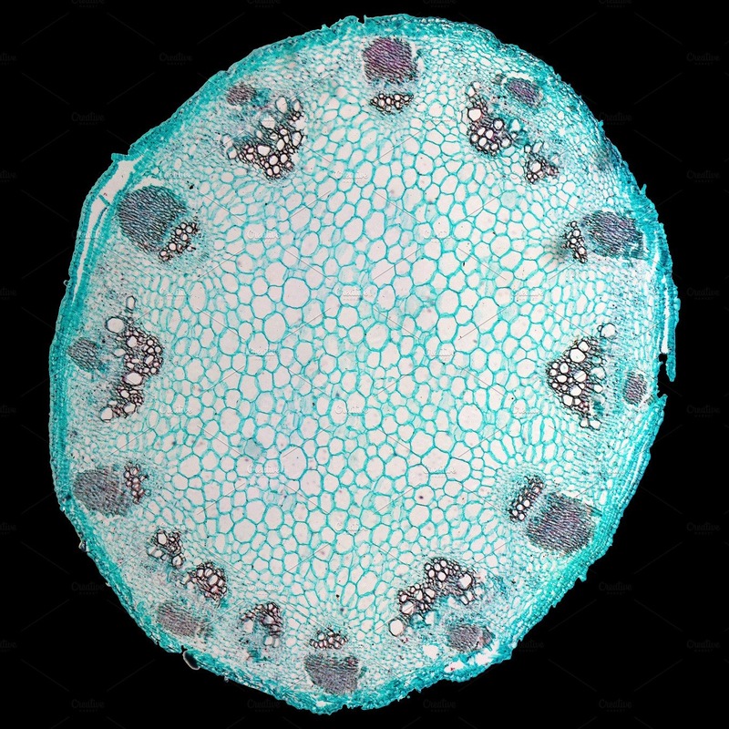
- Stereomicroscopes: Also known as dissecting microscopes, they provide a 3D view. They are ideal for examining the surfaces of plant samples or the overall structure of a cell.
- Confocal Microscopes: These enable scientists to construct a three-dimensional image of a plant cell. Confocal microscopes are particularly useful for viewing cellular activities in real time.
- Electron Microscopes: These offer the highest level of magnification. Electron microscopes are essential for seeing molecular details, like the intricacies of the cell wall.
Each type of microscope plays a critical role in expanding our knowledge of plant cells. They are key to uncovering the secrets held deep within plant tissues.
Preparing Plant Cell Samples for Observation
Before we can gaze into the micro-world of a plant cell under a microscope, we need to prepare the samples. Proper preparation is vital to ensure a clear view of the cell structures. It involves a series of careful steps that must be done with precision to avoid damaging the delicate samples.
Steps for Staining and Mounting Plant Cells
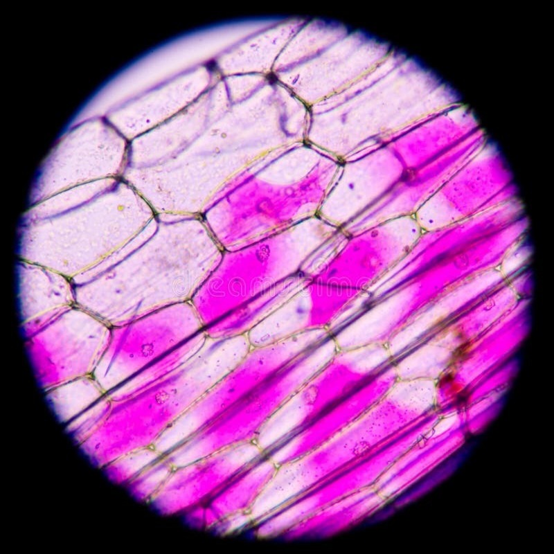
Observing the diverse components of a plant cell under a microscope is easier when the cells are stained. Staining adds contrast, making the intricate parts visible. Here’s how to do it:
- Select Fresh Samples: Choose newly-grown leaves or roots for dynamic cellular activity.
- Slice Thinly: Cut the sample in thin sections to allow light to pass through.
- Fix Cells: Use chemical fixatives to preserve cellular structures.
- Rinse Thoroughly: Clear away any excess fixative to avoid obscuring the view.
- Apply Stain: Choose the appropriate dye to highlight specific cell parts.
- Rinse Again: Remove surplus stain to improve clarity.
- Mount on Slides: Carefully place the stained sample on a microscope slide.
- Cover with Care: Seal the sample with a cover slip, avoiding air bubbles.
Each step in the preparation process has a purpose. Cutting the sample thinly allows detailed observation. Using fixatives keeps the plant cells from breaking down. Staining brings cellular components into sharp relief. Proper rinsing and mounting preserver the integrity of the cell’s appearance. It’s a meticulous task, but the reward is a window into the hidden world of plant cells.
Key Structures of Plant Cells Under the Microscope
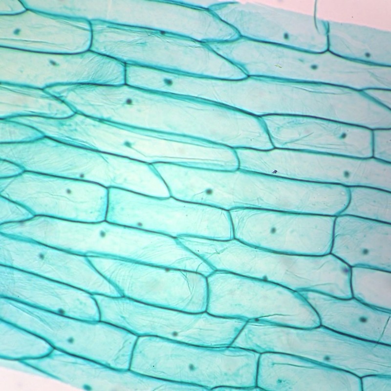
When gazing at a plant cell under a microscope, various key structures come into focus. These parts are vital for the cell’s function and overall health of the plant. Let’s explore some of these essential components.
Understanding the Role of the Cell Wall and Membranes
The cell wall is a plant cell’s outer layer providing support and structure. It protects the cell from outside forces and gives shape to the plant. Right inside, the cell membrane controls the movement of substances in and out. This gatekeeping is crucial for maintaining the cell’s internal balance.
Chloroplasts and Photosynthesis
Chloroplasts are the green energy factories of plant cells. They absorb sunlight and turn it into energy, enabling photosynthesis. This process is essential for plant growth and sustenance.
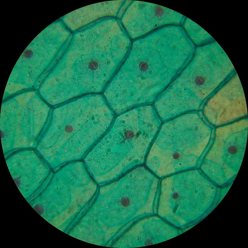
Nucleus and DNA: The Control Center
The nucleus is the command center of the plant cell. It houses DNA, the blueprint for all cellular activities. Monitoring the nucleus under a microscope can reveal a lot about a plant’s health and genetic potential.
Vacuoles and Storage
Vacuoles serve as storage bins inside plant cells. They hold various substances such as nutrients, water, and waste. Their size can tell us a lot about the cell’s current state and needs.
Techniques for Capturing Detailed Images
Capturing the subtle nuances of plant cells requires advanced techniques. Researchers and scientists use various methods to create clear, detailed images of these microscopic structures. These images provide valuable insights into the cell’s behavior and physiology. Now, let’s delve into one of the most revealing techniques used in plant cell microscopy.
Time-Lapse Microscopy to Observe Cell Processes
Time-lapse microscopy is a powerful tool for observing plant cells under a microscope. It records a series of images over time, capturing cell processes as they happen. Here’s how it works:
- Set Up the Microscope: Ensure the equipment is properly calibrated for time-lapse imaging.
- Prepare the Sample: Follow the steps for staining and mounting plant cells to enhance visibility.
- Start Recording: Begin capturing images at regular intervals. This could be seconds, minutes, or hours apart.
- Compile Images: Combine the individual images into a sequence that shows cell changes over time.
By using time-lapse microscopy, scientists can watch how a plant cell grows, divides, or responds to stimuli. This real-time view is crucial for understanding plant health and development. The technique offers a dynamic look at processes like photosynthesis, cell wall formation, and more. It sheds light on the life of a plant cell in ways static images cannot. Time-lapse microscopy unveils the beautiful symphony of life at the cellular level.
Interpreting Microscopic Data
When observing a plant cell under a microscope, interpretation is as crucial as the observation itself. Scientists spend hours meticulously analyzing microscopic data to understand the health and condition of plant cells. This analysis informs us about the growth and any potential issues that plants may face.
Analyzing Cell Morphology
Cell morphology refers to the shape, size, and structure of the cells. Under a microscope, experts look for the uniformity of cell walls and the arrangement of internal components. They check for the even distribution of chloroplasts which can indicate efficient photosynthesis. The nucleus’s appearance can also suggest how active the cell is in producing new proteins and plant tissues. By examining cell morphology, researchers can gather information about the plant’s growth patterns and overall vitality.
Identifying Signs of Plant Stress or Disease
Microscopes not only reveal detailed structures but also signs of distress within plant cells. Changes in cell morphology can signal stress due to environmental factors, such as drought or overexposure to sunlight. Discoloration in cells or the presence of abnormal structures often point to disease. Scientists also look for irregularities in vacuole size, which could mean nutritional deficiencies or toxin buildup. Identifying these signs early can lead to quicker responses in plant care and disease management, protecting crops and natural plant populations.
Advancements in Microscopic Technology
The field of microscopic technology has seen remarkable advancements. These improvements have greatly enhanced our ability to observe a plant cell under a microscope. Researchers now have access to sharper, more detailed images than ever before.
The Evolution of High-Resolution Plant Cell Imaging
High-resolution imaging technology has transformed our study of plant cells. Innovations like improved lenses and digital software provide clearer images. This has allowed us to view cellular activities with astounding precision.
- Enhanced Optical Microscopes: Recent upgrades have improved magnification and resolution.
- Digital Imaging Techniques: Software now aids in capturing and analyzing cell images.
- Fluorescence Microscopy: This method lights up specific cell parts, aiding in their identification.
- Atomic Force Microscopy: We can now observe the surface of cells at the atomic level.
These advancements have given us new perspectives. They enable scientists to uncover the tiny secrets within plant cells. This has a big impact on our knowledge of plant biology and its applications.
Conclusion
As we have explored the microscopic world of plant cells, we see that microscopy has opened up vast areas of research and knowledge. The journey from the basic compound microscope to the advanced digital imaging techniques has significantly impacted our understanding of plant biology. Looking ahead, the future of plant cell research and microscopy promises to bring even more exciting discoveries and innovations.
The Future of Plant Cell Research and Microscopy
The continual advancement in microscopic technology will undoubtedly enhance our observation of plant cells under a microscope. With AI integration, image analysis will become quicker and more accurate, facilitating the study of cell processes in real time. Nanotechnology may introduce new staining methods, allowing for non-invasive tracking of cellular functions. Moreover, advancements in 3D imaging could provide a more holistic view of cell interactions within the plant tissue. As we forge ahead, the merging of microscopy with other biotechnological tools is set to revolutionize the field of plant science. This evolution will further our knowledge, aid in food security, and improve ecological sustainability. The secrets that have long eluded us are nearing revelation as we continue to zoom in on the hidden universe of plant cells.


