The Role of Microscopy in Cell Observation
Microscopy is a pivotal tool in biology and medicine. It allows us to peer into the intricate world of cells under microscope. With microscopy, we can explore cell structures invisible to the naked eye. Let’s delve into how this is achieved.
Understanding the Basics
At its core, microscopy uses lenses to magnify images of tiny objects. Cells are prime subjects. Lenses enlarge the view, so we can observe cells in detail.
Magnification and Resolution
Two key factors define microscopy’s effectiveness: magnification and resolution. Magnification enlarges the cell image. Resolution clarifies it. Together, they bring cells into sharp focus for observers.
Advances in Microscopic Techniques
Over time, innovations have advanced microscopy. Modern techniques reveal more about cells under microscope than ever before. They allow researchers to see cell components and understand their functions.
Importance for Research and Diagnosis
Scientists use microscopy to study cell biology. This research is vital for understanding diseases. In medicine, microscopy helps diagnose conditions based on cellular changes. It truly is an indispensable tool in labs across the globe.
Microscopy in Education
Microscopes also play a central role in education. They help students see cells firsthand. This visual access is vital for learning about cell biology. It lays a foundation for future scientific exploration.
Microscopy remains a fundamental method for observing cells under microscope. It enables discoveries and aids in the fight against diseases. It is not just a window into the hidden world of cells; it’s the key that unlocks the door.
Types of Microscopes Used to Study Cells
When exploring the hidden world of cells under microscope, the type of microscope used is crucial. Various microscopes offer different levels of detail, accessibility, and functionality. Here are the primary microscopes employed in the study of cells:
- Compound Microscopes: These are the most common type. They use two sets of lenses for higher magnification and are ideal for observing thin slices of cells.
- Stereomicroscopes (also known as dissecting microscopes): With a lower magnification, these are perfect for viewing larger, 3D specimens, such as tissue samples.
- Confocal Microscopes: Employing laser light to scan cells, these microscopes produce high-resolution images. They are particularly useful in viewing the fine details within a cell.
- Electron Microscopes: These fall into two categories, Transmission Electron Microscopes (TEM) and Scanning Electron Microscopes (SEM). TEMs provide detailed internal cellular views, while SEMs focus on surface details.
- Fluorescence Microscopes: These use specific wavelengths of light to illuminate proteins or other molecules within cells, often tagged with fluorescent markers.
Choosing the right microscope depends on the study subject and the level of detail required. Scientists using these tools can discover the intricate details and processes that define life at the cellular level. By employing various types of microscopes, researchers can learn more than ever before about cells under microscope, helping them to unlock the mysteries of biology and medicine.
Preparing Biological Samples for Microscopic Analysis
Before cells under microscope can reveal their secrets, they must be carefully prepared. Proper sample preparation is critical to obtaining clear, accurate microscopic images. Here’s how scientists prepare biological samples for microscopic analysis:
- Cleaning: Samples must be free of contaminants. This includes dust, oils, and any unwanted particles.
- Fixation: Cells or tissues are treated with chemicals to preserve their structures. This step is essential to prevent degradation.
- Sectioning: For thicker samples, thin slices called sections are needed. They allow light or electrons to pass through for better visualization.
- Mounting: The prepared sections are placed on slides. Slides provide a stable platform for observation under the microscope.
- Drying: Removing moisture prevents the distortion of cell structures. Careful drying helps maintain the integrity of the samples.
Each step is done with precision to ensure that the cells under microscope will be displayed in their truest form. Whether for educational purposes or advanced research, proper sample preparation lays the groundwork for successful microscopic study. Through these meticulous methods, the minute details of cells are brought into sharp focus, enabling scientists and students alike to explore the complexities of life at a microscopic level.
The Cell Theory and Its Impact on Microscopy
The cell theory has been a cornerstone in biological sciences. It states that all living things are made up of cells, cells are the basic units of structure and function in organisms, and new cells are produced from existing cells. This theory has had profound implications for microscopy and our understanding of cellular structures.
Guiding Microscopic Research
The acceptance of the cell theory shifted the focus of microscopic research. Scientists began systematically studying cells as fundamental living units. It drove advancements in microscope design and techniques, aiming to better observe and understand cell functions.
Improving Microscopic Techniques
As the theory emphasized cells’ pivotal role in life, the demand for higher resolution and magnification in microscopes grew. This led to refinements in lens crafting and the introduction of electron microscopy, which brought forth unprecedented detail of cells under microscope.
Influencing Educational Frameworks
Cell theory’s impact extends into education. It has shaped curricula, where students now learn cell structure and function with hands-on microscopy. Such education has advanced the literacy of students in biology and empowered future researchers.
The cell theory, by asserting the universality of cells in living organisms, has been fundamental in driving the development and use of microscopy. It supports why cells under microscope remain a subject of fascination and study across all levels of biological research and education.
Common Cell Structures Visible Under a Microscope
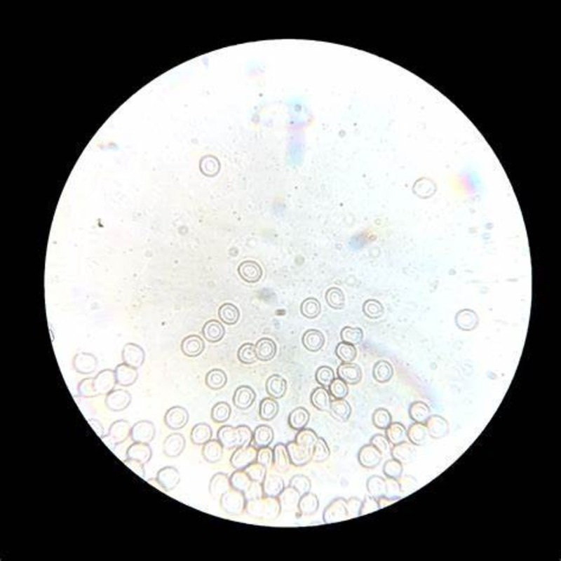
When we place cells under microscope, we see an array of structures unique to each cell type. By using various microscopy techniques, scientists can identify different parts that make up a cell. Here are several common structures often observed:
- Nucleus: This is the cell’s command center containing genetic material. It’s typically the most prominent structure in eukaryotic cells.
- Mitochondria: Known as the powerhouse of the cell, mitochondria are visible as rod-like structures. They are the sites of energy production.
- Cell Membrane: The thin layer enclosing the cell’s contents, this membrane controls what enters and exits the cell.
- Cytoplasm: This gel-like substance fills the cell and holds organelles in place. It’s where many cellular activities occur.
Each of these structures plays a critical role in the life of a cell. Viewing cells under microscope allows students and researchers to gain a deeper understanding of cellular function and complexity.
Staining Techniques: Enhancing Cellular Contrast
To reveal the intricate details of cells under microscope, staining is critical. Staining techniques apply colors to cellular components. This makes structures stand out against the background. Cells, in their natural state, often lack contrast. With stains, we can see them more clearly.
Here are some common staining methods used in microscopy:
- Simple Stains: These use one dye to color cells. They help identify shape and size.
- Differential Stains: This method uses two or more dyes. It distinguishes different cell types or parts.
- Fluorescent Stains: These attach to specific molecules. Under UV light, they glow, showing precise structures.
- Immunostaining: This technique uses antibodies. Antibodies bind to target molecules, which are then visualized with dyes.
Each technique serves a unique purpose. For example, simple stains might show the overall cell shape. Differential stains could highlight the nucleus or bacteria types. Fluorescent stains are used to see proteins or DNA. Immunostaining is ideal for pinpointing specific molecules.
Choosing the right stain depends on the inquiry. With the right staining, cells under microscope can display their hidden details. This enhances our understanding of cellular biology. Students and researchers rely on staining to study cells closely. It bridges the gap between the unseen microscopic world and our ability to perceive it.
Live Cell Imaging: Observing Cellular Processes in Real Time
In the fascinating study of cells under microscope, live cell imaging stands out. It lets us watch cells live, in their natural, bustling state. Here’s why it’s pivotal in cell biology.
- Real-time monitoring: Live cell imaging captures cellular events as they unfold. This is vital for understanding the dynamics of cell function.
- Interaction observation: Researchers see how cells interact with their environment. This helps to decipher how they respond to stimuli.
- Cellular lifecycle study: The tool allows us to track the life cycle of a cell. We witness everything from division to death.
- Drug effects: It shows how cells react to medical treatments. This is crucial for developing new drugs.
- Movement tracking: Live imaging captures how cells move. Migration and motility are key in processes like wound healing.
Advanced microscopes with time-lapse features are used for live cell imaging. These devices minimize light exposure to prevent cell harm. They keep cells healthy during prolonged observations.
The insights gained from live cell imaging are immense. They contribute to our knowledge of cellular processes. This method sheds light on the inner life of cells under microscope. As a result, it advances both research and medicine. This technique showcases the cell’s story, minute by minute.
Ethical Considerations in Microscopic Research of Cells
When examining cells under microscope, ethical considerations are crucial. Researchers must navigate a complex landscape of morality and regulations. Here are primary ethical aspects they face:
- Consent: For human tissue samples, informed consent is key.
- Privacy: Personal information linked to cell samples must be protected. Researchers ensure confidentiality to maintain donor trust.
- Animal welfare: Studies involving animal cells must minimize harm. This follows the ‘Three Rs’: Replacement, Reduction, and Refinement of animal use.
- Biosafety: Handling cells, especially pathogens, needs extreme care. Labs follow strict safety protocols to prevent contamination and spread.
- Intellectual property: Research findings, including discoveries made through microscopy, can raise questions about ownership and sharing of information.
- Publication ethics: Presenting research results must be truthful and clear. This avoids misleading the scientific community or public.
Through attention to these areas, the scientific community ensures that research involving cells under microscope is conducted responsibly. It balances the quest for knowledge with respect for living organisms and ethical norms. This approach fosters trust in science and supports the advancement of medicine and biology.


