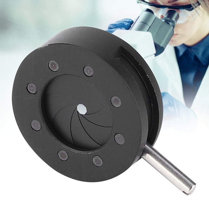
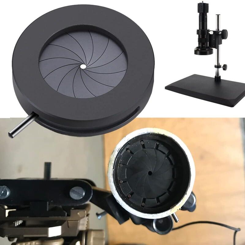
Introduction to Iris Diaphragm in Microscopy
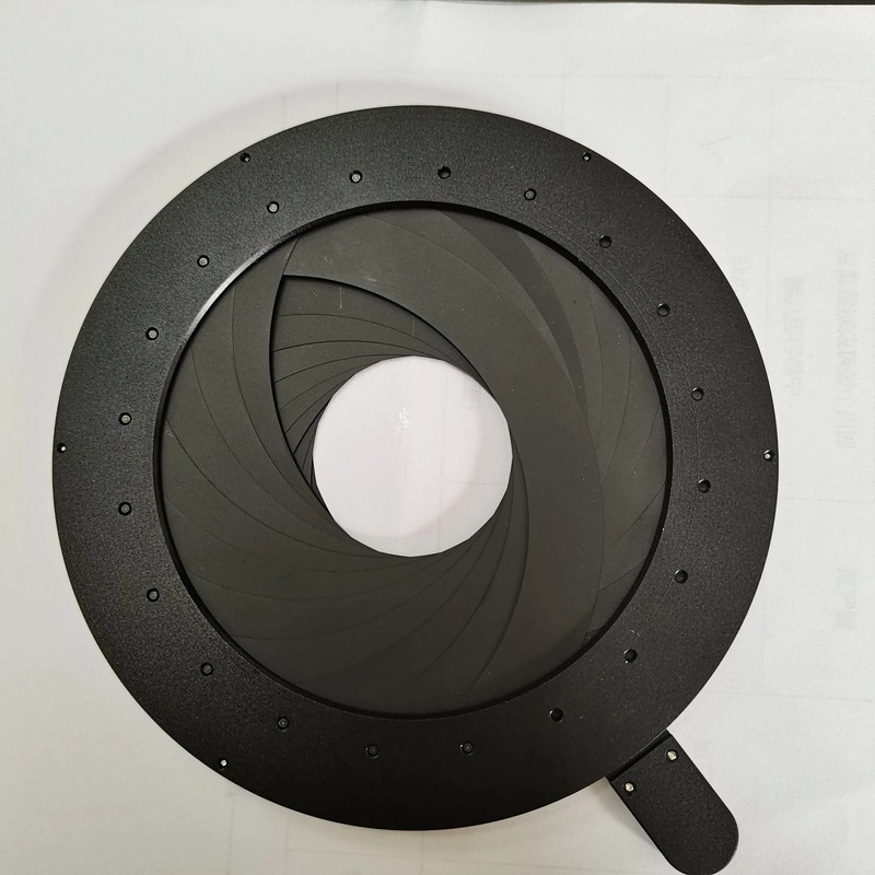
The iris diaphragm is a crucial component of a microscope. It sits beneath the stage and controls light. With an adjustable aperture, it influences the intensity and diameter of light reaching the specimen. This part resembles the eye’s iris, hence the name. It is commonly found in compound microscopes but may also appear in other types.
Understanding how to use the iris diaphragm is key for microscopists. Adjusting it alters the contrast and resolution of the microscopic image. It can sharpen the focus or highlight certain details. An open diaphragm lets in more light, making an image brighter but possibly washing out details. A closed diaphragm reduces light, enhancing contrast but can make an image too dark.
Proper manipulation of the iris diaphragm contributes greatly to image quality. Thus, it’s essential for both beginners and seasoned users to understand its role. We will delve into detailed functions and maintenance tips to ensure effective use of the iris diaphragm microscope feature in further sections.
The Function of the Iris Diaphragm
The iris diaphragm microscope feature serves a pivotal function. It helps to regulate light that illuminates the sample being observed. By changing the size of the aperture, it permits more or less light through. This affects the brilliance and clarity of the image seen through the microscope’s eyepiece. Precise control of the iris diaphragm is essential for various microscope techniques. These techniques include bright field, dark field, and phase contrast microscopy.
When using the iris diaphragm, microscopists can manage the quality of the image. A smaller aperture increases contrast, making structural details in the specimen more distinct. Conversely, a larger aperture can make the field of view brighter but may reduce contrast. The correct setting of the iris diaphragm can reveal fine details or focus on specific aspects of the specimen.
Beyond simply altering the brightness and contrast, the iris diaphragm influences the depth of field. Depth of field refers to how much of the vertical space in the specimen is in clear focus at one time. A narrow aperture enhances the depth of field, allowing more of the specimen to appear in focus. This is particularly useful for thicker specimens where layers of interest exist at different depths.
The ability to adjust the iris diaphragm according to the specimen’s requirements is a skill that microscopists must hone. Whether it’s for studying cell structures, microorganisms, or tissue samples, the iris diaphragm aids in achieving the best possible microscopic examination. It is an indispensable tool for scientists and researchers in the fields of biology, material science, and many other areas where microscopy plays a fundamental role.
Adjusting Contrast and Resolution with the Iris Diaphragm
Achieving the right contrast and resolution in microscopy is critical for clear observation. The iris diaphragm microscope feature is central to this process. It does more than just control the amount of light. It finely tunes the visual aspects of the specimen under study.
Here’s how to adjust contrast with the iris diaphragm:
- Close the iris diaphragm slightly to enhance the contrast. By limiting light, it makes specimen details more prominent against the background.
- Observe the effect on the sample. If the image is too dark, open the iris diaphragm a little.
- Find balance. The aim is to have clearly defined details without losing visibility due to darkness.
Resolution adjustment using the iris diaphragm involves the following steps:
- Start with the iris diaphragm fully open to see the maximum field of view.
- Gradually close the diaphragm to sharpen the focus on the specimen’s details.
- Avoid over-closing, which can reduce image quality.
- Stop when the highest resolution is achieved without compromising brightness.
Experimenting with the iris diaphragm settings is the best way to understand its effect on contrast and resolution. Practicing this regularly will refine your microscopy skills, enhancing the visualization of specimen details. Remember, the goal is to achieve a balance that results in crisp, clear images that reveal the finest structures of what you’re examining.

The Impact of Iris Diaphragm on Depth of Field
The iris diaphragm microscope component shapes the depth of field in view. It lets users focus on different layers within a specimen. A closed iris diaphragm increases the depth of field, meaning more of the subject is in clear focus. This setting is game-changing for viewing complex structures or thick samples.
Here’s a closer look at how the iris diaphragm affects depth of field:
- Narrow Aperture: A small opening provides a greater depth of field. You’ll see more detail at various depths at once.
- Wide Aperture: A large opening gives a shallower depth of field. It focuses on a specific plane within the sample.
- Adjustment: By adjusting the aperture, you alter the depth of focus. This helps to pinpoint areas of interest in your specimen.
To make the most of the depth of field, you’ll need to practice adjusting the iris diaphragm. Start with a wide aperture and then narrow it down slowly. Do this until you find the perfect focus for your needs. Each adjustment can bring out details that weren’t visible before.
The depth of field is critical for microscopists. It can mean the difference between a clear or a blurry observation. With the iris diaphragm microscope feature, you have control over this essential aspect of microscopy.
Operating the Iris Diaphragm for Optimal Imaging
Operating the iris diaphragm correctly is key to optimal imaging in microscopy. Here are steps to adjust the iris diaphragm:
- Start with the iris diaphragm wide open. This will flood the field of view with light.
- Slowly close the diaphragm while observing the specimen. Notice changes in brightness and sharpness.
- Continue to adjust until you achieve a balanced image. The goal is clear details without excess brightness.
Keep in mind that different specimens may need different settings. Here are some tips for various applications:
- For thin specimens, a slightly more open iris diaphragm can provide enough contrast.
- For thick or layered samples, a narrower aperture increases depth of field and detail clarity.
- In low-light conditions, open the diaphragm more to brighten the image.
Remember, the iris diaphragm microscope setting is not a one-size-fits-all. Practicing adjustments will help refine your technique. Always aim for the balance that gives you the clearest and most detailed view of your specimen.
Common Misconceptions About the Iris Diaphragm
The iris diaphragm microscope feature often comes with its fair share of myths. These misconceptions could impact the proper use and understanding of this critical component. Let’s debunk some common myths and shed some light on the facts.
- Myth: The iris diaphragm should always be fully open for the best clarity. Fact: Opening the diaphragm fully can sometimes reduce contrast, making details less visible.
- Myth: It’s best to keep the diaphragm closed for all types of samples. Fact: This setting can darken the image too much, hiding fine details. Different specimens require varying levels of light and contrast.
- Myth: Adjusting the iris diaphragm doesn’t affect image resolution. Fact: The iris diaphragm plays a significant role in fine-tuning both the contrast and the sharpness of the image, thereby influencing resolution.
- Myth: The iris diaphragm is only useful for high magnification. Fact: This tool is valuable across various magnifications. It helps reveal intricate details at lower magnifications too.
- Myth: Use of the iris diaphragm is intuitive and doesn’t require practice. Fact: Mastering the proper use of an iris diaphragm microscope feature takes time and experimentation.
- Myth: There’s a standard setting for the iris diaphragm that works for every situation. Fact: Ideal diaphragm settings depend on the specimen and lighting conditions. They require careful, situation-specific adjustments.
Understanding the real capabilities of the iris diaphragm enhances microscopy techniques. As you work with various specimens, it’s important to approach each with a clear mind, free from these myths.
Maintenance and Care for Microscope Iris Diaphragms
Proper maintenance of the iris diaphragm is essential to keep a microscope in good working order. Here are some straightforward tips to ensure your iris diaphragm microscope component remains in top condition:
- Regularly check the diaphragm for debris and dust. Clean gently with a soft brush or compressed air.
- Use lens paper moistened with distilled water or a mild cleaning solution to wipe the blades. Do so with great care to avoid damage.
- Avoid touching the iris diaphragm with fingers. Oils from the skin can corrode the metal parts.
- Ensure that the diaphragm moves smoothly. If it sticks, do not force it. Consult the microscope manual or a professional for advice.
- Store the microscope in a dry, cool place. This helps prevent mold and rust from forming on the diaphragm.
- Have the microscope serviced regularly. This includes a professional cleaning and lubrication of moving parts.
- Do not disassemble the iris diaphragm. If it seems faulty, seek professional repair services.
By taking simple steps in maintenance, you can extend the functional life of your iris diaphragm microscope component. This, in turn, ensures that you continue to get high-quality images from your microscopy work.
Advances in Iris Diaphragm Design and Technology
The design and technology of the iris diaphragm microscope component have seen notable advancements. These improvements aim to enhance microscopy. They make the operation of the iris diaphragm smoother and more precise.
Developers have introduced materials that resist corrosion and wear. This leads to longer-lasting diaphragms. They have made strides in the precision of the aperture control. Users can make finer adjustments than before. There is now better control over light and contrast.
Modern iris diaphragms also feature designs that are easier to clean. This minimizes maintenance needs. Some models now come with anti-reflective coatings. These reduce glare and improve image quality.
New technologies integrate computer controls with the iris diaphragm. This allows for automatic adjustments during imaging. Researchers can focus on the sample instead of manual settings.
In summary, the iris diaphragm microscope component continues to evolve. These advancements make microscopy more effective. They provide clearer images and easier handling, helping researchers in their work.


