Introduction to Smooth Muscle Tissue
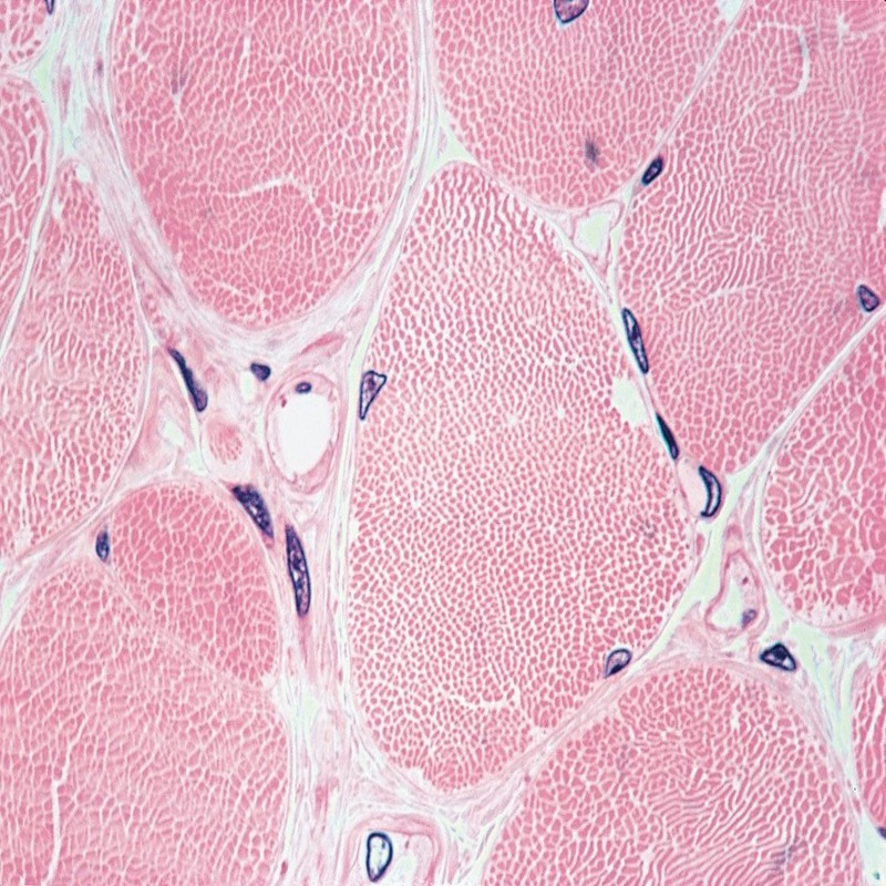
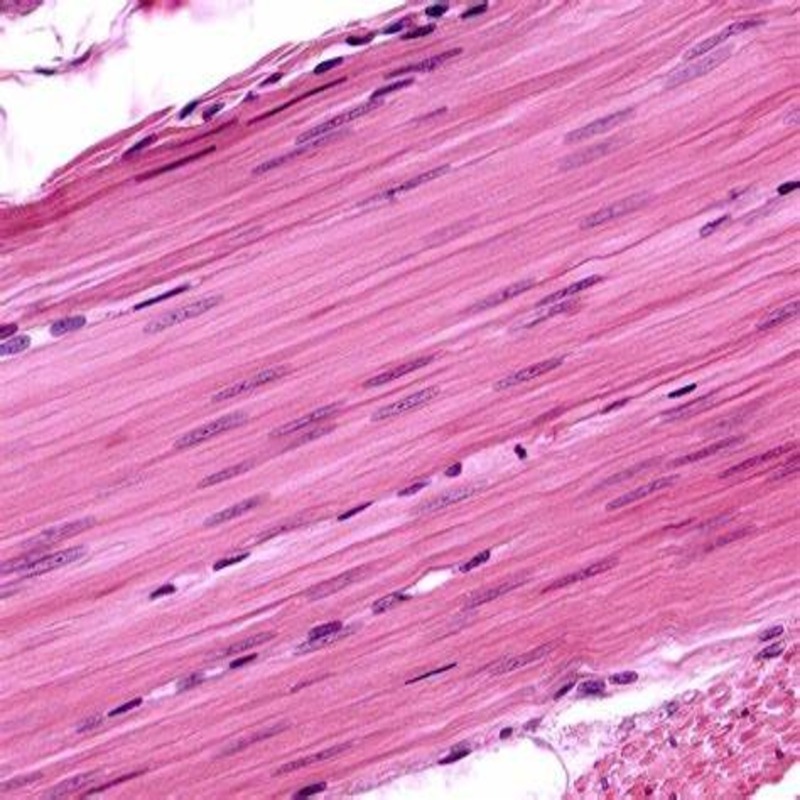
Smooth muscle tissue is a key part of our body. It is found in the walls of our internal organs. For example, it is in our stomach, bladder, and blood vessels. This tissue helps control many functions. It contracts and relaxes to aid in digestion, blood flow, and more. Unlike skeletal muscle, smooth muscle is not under our conscious control. It works on its own without our thought. Under the microscope, smooth muscle looks different than other muscles. The cells are spindle-shaped, and they have a single nucleus in the center. They connect in a network that lets them work in sync. This unique structure is why they can push things through our organs.
Research has shown that these muscles play a big role in our health. If they change or get sick, it can lead to serious issues. It is why scientists study smooth muscle cells in detail. With new tech in 2025, we can now see these cells in ways we could not before. This blog post will talk about the latest tools we use to study smooth muscle under the microscope. We will also discuss how these advances help us learn more about their role in our body.
Advances in Microscopic Techniques in 2025
The field of microscopy has seen significant advancements in 2025. These progressions allow a more detailed study of smooth muscle under microscope. Here are the key developments that have been pivotal:
- Enhanced Resolution: The new microscopes boast an increased resolution. This upgrade offers clearer images of smooth muscle tissue. Researchers can now observe finer structural details.
- High-Speed Imaging: Advances in high-speed imaging enable the capture of smooth muscle movements. Scientists can watch how these muscles contract and relax in real-time.
- 3D Imaging Techniques: Cutting-edge 3D imaging allows a deeper look into the arrangement of smooth muscle cells. It helps in understanding how these cells form networks.
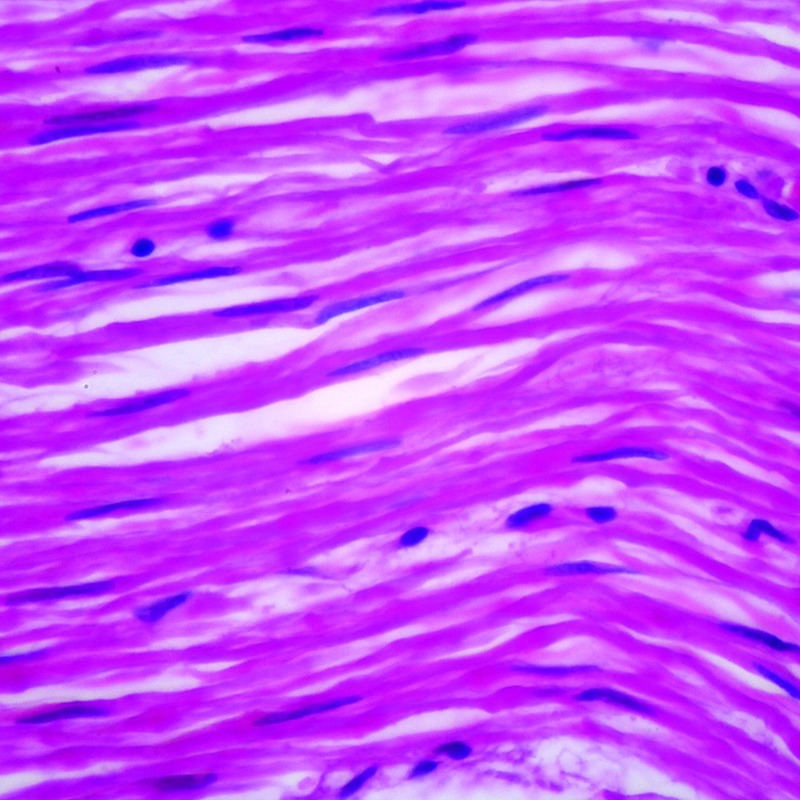
- Improved Staining Methods: Novel staining methods distinguish smooth muscle cells more effectively. This results in better contrast and visibility under the microscope.
- Automation In Image Analysis: With improved software, image analysis becomes faster and more accurate. It helps in the quantification of smooth muscle characteristics.
- Accessibility of Advanced Tools: Lastly, these tools are becoming more accessible to labs around the world. This means more scientists can study the nuanced behavior of smooth muscle.
Thanks to these advances, our comprehension of smooth muscle microstructure is advancing rapidly. They are changing how we look at the details of these critical tissues.
Key Characteristics of Smooth Muscle Cells
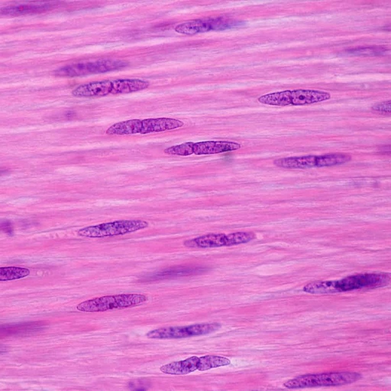
Smooth muscle cells have distinct features that set them apart. Under the microscope, these cells show up as spindle-shaped with pointed ends. This shape is unlike the cylindrical form seen in skeletal muscle. Each cell hosts a single, centrally located nucleus. This is a contrast from the multiple nuclei found in other muscle types.
Researchers observe these cells working together in sheets or layers. They link via gap junctions, allowing them to contract as one unit. This connectivity is crucial for their function in organs. The cells’ ability to stretch and maintain tension is key. It gives organs like the bladder the capacity to hold fluids.
When viewing smooth muscle under microscope, scientists note the absence of striations. This is a major difference from skeletal and cardiac muscle, which both have striped patterns. The lack of striations is due to the arrangement of protein fibers within the cells. These fibers slide past each other to make contraction possible.
The structure of smooth muscle cells helps them to work for long periods without tiring. This endurance is because they contract slower than skeletal muscles. But, this slow contraction is also very powerful, ideal for the necessary functions these muscles perform.
Understanding these characteristics of smooth muscle cells is vital. It aids in grasping their unique role in our body systems. Advances in microscopy have been instrumental in deepening this understanding.
Smooth Muscle vs. Skeletal and Cardiac Muscle
When we compare smooth muscle to skeletal and cardiac muscles, a few key differences stand out. First, in terms of control, smooth muscle operates involuntarily. This means our bodies run these muscles without our conscious effort. In contrast, skeletal muscle moves when we decide to move, like when we walk or lift something. Cardiac muscle, similar to smooth muscle, works on its own, keeping our heart beating without thought.
Another major difference lies in appearance. As we’ve discussed, smooth muscle cells are spindle-shaped and lack striations. In skeletal muscle, the cells are long, cylindrical, and feature a striped pattern due to their protein fiber alignment. Cardiac muscle also has striations, but the cells connect at branching points creating a unique web-like design.
Functionally, smooth muscle cells are the marathon runners. They contract slowly but they can go on for a long time. This endurance is perfect for their role in our bodies, managing continuous tasks like controlling blood pressure and moving food through the digestive system. Skeletal muscles are sprinters; they contract quickly but tire easily. Cardiac muscle sits in between, contracting rhythmically and powerfully to keep blood circulating.
Each muscle type is adapted to its specific role. Smooth muscle ensures the steady functioning of our organs. Skeletal muscles handle rapid, forceful movements. Cardiac muscle, on the other hand, is specialized for never-ending and rhythmic contractions. By studying smooth muscle under a microscope, we get clearer insights into these differences and their importance in human physiology.
Investigating Smooth Muscle in Various Organs
When we study smooth muscle under microscope, we find it in many organs. It plays unique roles across the body. Let’s delve into some places where smooth muscle does its work:
- Digestive System: In the gut, smooth muscle cells line the walls. They help in moving food through digestion. This is called peristalsis.
- Respiratory System: These muscles wrap around airways in the lungs. They control the flow of air by tightening or relaxing.
- Vascular System: Smooth muscle in blood vessel walls helps control blood pressure. When these muscles contract, they change the vessel diameter.
- Urinary System: The bladder is made up of smooth muscle. It allows the bladder to store and release urine.
- Reproductive System: In females, smooth muscle helps in the movement of eggs. In males, it plays a role in the flow of semen.
Researchers can now see how these cells work in different settings, thanks to new microscopic techniques. For instance, the high-speed imaging captures the muscle’s movement in the bladder or gut. The improved staining methods show the muscle’s details in the blood vessels.
These studies reveal how smooth muscle maintains health in different organs. They also show what may happen when something goes wrong. For example, if the muscle in the blood vessels cannot work right, it may lead to high blood pressure.
By taking a closer look at smooth muscle in these organs, scientists can better understand diseases. They can see changes in the muscle’s structure or function with the microscope’s improved capabilities. This knowledge could lead to new ways to treat organ-specific conditions involving smooth muscle.
The Role of Smooth Muscle in Human Physiology
The role of smooth muscle in human physiology cannot be overstated. It plays a crucial part in the everyday operations of our body systems. Understanding its functions deepens with each new observation under the microscope.
Smooth muscle is central in managing vital involuntary processes. This includes the movement of food through our digestive tract, which is essential for nutrient absorption. In the respiratory system, smooth muscle helps regulate our breathing by adjusting the airway diameter. This action is automatic and adjusts to our body’s oxygen needs.
Blood pressure regulation also hinges on the performance of smooth muscle. These muscles flexibly contract or relax to alter the diameter of blood vessels. By doing so, smooth muscles facilitate the control of blood flow and pressure throughout the body.
In the urinary system, smooth muscle is just as crucial. The bladder relies on it to store or dispel urine efficiently. As for the reproductive system, this muscle type assists in the passage of gametes, affecting fertility and reproduction.
Smooth muscle also maintains tension and elasticity in various organs. This quality is indispensable, as it allows structures like the stomach and intestines to return to their original shape after stretching.
By studying smooth muscle under a microscope, researchers have gathered that its seamless function is integral. It is a key player in homeostasis, maintaining an internal balance within the body.
To sum it up, smooth muscle is a workhorse in human physiology. Its involuntarily controlled actions support core bodily functions that sustain life. Advanced microscopy has revealed the intricate operations of these muscles. This aids in our overall understanding of how our bodies function at a cellular level.
Pathological Changes in Smooth Muscle Observed Under Microscope
When studying smooth muscle under microscope, researchers also focus on abnormalities. These can point to various diseases and health issues. Microscopy has made it possible to spot problems in the muscle’s structure and behavior. Here’s what scientists often look for:
- Irregular Shape: Smooth muscle cells in good health are spindle-shaped. If they appear irregular or misshapen, it can suggest a problem.
- Nucleus Alterations: Changes in the size or number of nuclei within these cells are red flags. They might indicate cellular stress or damage.
- Disorganized Arrangement: Normally, these cells form orderly networks. A chaotic or disordered arrangement could mean that there’s tissue dysfunction.
- Altered Contraction: Studying how these muscles move is critical. If there’s a change in their contraction patterns, it might be a sign of disease.
- Fibrosis: The presence of excess connective tissue, or fibrosis, can be detected. This suggests that the smooth muscle is overworking or repairing itself excessively.
Microscopes show us much more than just the muscle’s health. They help in disease diagnosis and treatment planning. For instance, changes seen under the microscope can lead to insights on high blood pressure or asthma.
When alterations are detected early, it’s often possible to treat the problem more effectively. That’s why the role of microscopes in researching smooth muscle is so vital. It allows for a deeper understanding not just of normal function, but also of the pathologies that affect human health.
Future of Smooth Muscle Research and Microscopy
The future of smooth muscle research looks bright and promising. As we continue advancing in microscopic techniques, our understanding of smooth muscle in human physiology is set to deepen. Here are the expected developments and their potential impacts:
- Increased Imaging Precision: We anticipate even higher resolution imaging. This will allow for the observation of subtler details within smooth muscle cells.
- Advanced Analytical Software: As we better analyze images, we will quickly identify abnormalities. This will aid in the early detection of diseases linked to smooth muscle.
- Wider Availability of Tools: Cutting-edge microscopes should become more common. This will enable more labs to contribute to smooth muscle research.
- Molecular-Level Insights: Future efforts may focus on examining smooth muscle at the molecular level. It could uncover new findings about muscle functionality and disorders.
- Interdisciplinary Research: Collaboration across fields like genetics and nanotechnology may lead to breakthroughs in our knowledge of smooth muscle.
- Innovative Treatment Approaches: By understanding the microstructure of smooth muscle, we might develop targeted therapies for related conditions.
As researchers employ these advanced microscopic tools to view smooth muscle under microscope, the potential for scientific discovery is vast. Every step forward in microscopy can lead to better treatments and a deeper understanding of crucial physiological processes.


