Introduction to Bacteria Observation
Exploring the microscopic world of bacteria is both fascinating and vital. Observing bacteria through a microscope is essential for scientific studies, medical diagnostics, and educational purposes. With the right tools and techniques, anyone can undertake the journey of discovering these minute life forms.
Importance of Microscope in Bacterial Study
To study bacteria, we need the amplifying power of a microscope. Bacteria are too small to see with the naked eye. Microscopes enable us to magnify them sufficiently to analyze their structures and behaviors. This magnification is crucial in research, diagnosis of diseases, and understanding the ecological role of bacteria.
Bacteria Size and Shapes
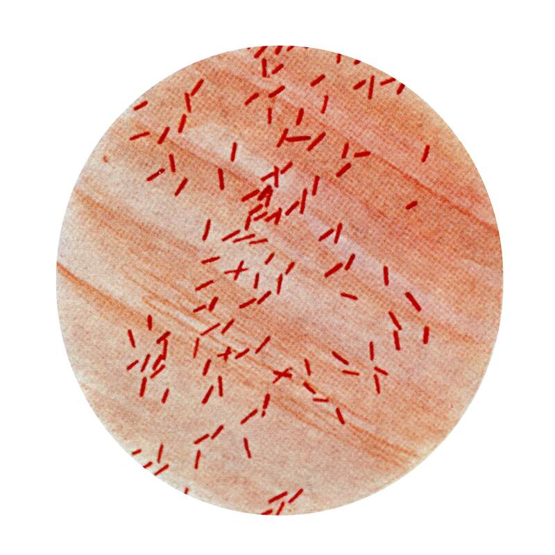
Bacteria exhibit a variety of shapes and sizes, usually measured in micrometers. Common shapes include spheres, rods, and spirals. These forms, not discernible without magnification, reveal a lot about bacterial functionality and classification when observed under a microscope.
Essential Microscope Equipment
To view bacteria, having the correct microscope equipment is essential. This equipment includes the microscope itself, along with slides, coverslips, and stains for sample preparation.
Types of Microscopes to Observe Bacteria
Primarily, two types of microscopes can view bacteria: compound microscopes and phase-contrast microscopes. Compound microscopes offer multiple magnifications and are very common. Phase-contrast microscopes enhance the contrast of bacteria without staining, preserving their natural state.
Preparing Your Microscope for Observation
Before observing bacteria, ensure the microscope is clean and on a stable surface. Plug it in or check batteries and adjust the light to a comfortable level. Gather all necessary materials, such as clean slides, stains, and a swab or loop for sample collection.
Sample Preparation Techniques
Preparing samples correctly is crucial for successful microscopy of bacteria.
Collecting Bacteria Samples
To begin, select an area where bacteria are likely present. Use a sterilized loop or swab to gather your specimen.
Staining: Enhancing Visibility
Apply a stain such as methylene blue to make bacteria more visible. Carefully drop the stain onto the sample and let it air dry.
Slide Mounting Basics
Place the stained sample on a slide, gently cover with a coverslip, and secure it to avoid slipping while observing.
Step-by-Step Microscopic Examination
Ensuring you conduct a thorough microscopic examination is key to identifying bacteria.
Starting with Low Magnification
Begin your observation with the microscope’s lowest magnification setting. This initial view allows you to focus on your sample and covers a larger area, making it easier to locate any bacterial presence. During this stage:
- Place your slide on the stage and secure it with stage clips.
- Use the 4x or 10x objective lens to get a broad view of the sample.
- Adjust the diaphragm to regulate light for clear visibility.
- Bring your sample into focus using the coarse adjustment knob.
Remember, low magnification gives you an overview and helps set up for detailed observation.
Increasing Magnification for Detailed View
Once focused on low power, switch to a higher magnification lens for an intricate look at the bacteria. The process involves:
- Moving to the 40x or 100x objective lens carefully.
- Using the fine adjustment knob to clarify the image.
- Adjusting the slide slightly as needed to keep the bacteria in view.
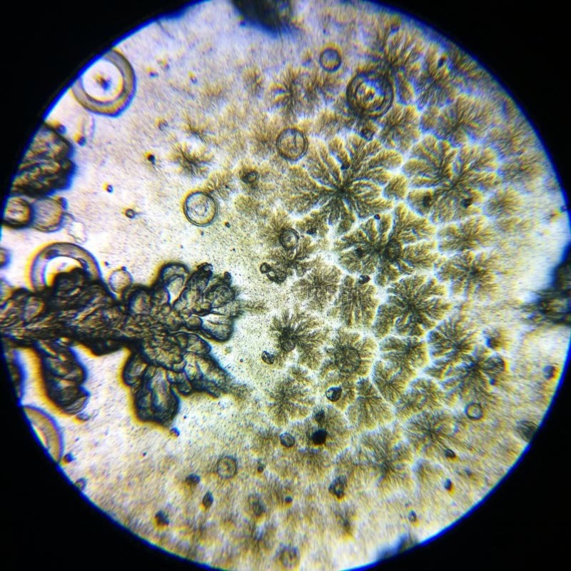
High magnification offers insight into the shape, size, and arrangement of bacteria, making them easier to identify.
Bacteria Identification Tips
To identify bacteria species, examine the distinct features:
- Note bacteria shapes — rods, spheres, or spirals.
- Observe how bacteria organize — in clusters, chains, or alone.
- Cross-check features with recognized patterns to identify species.
Utilize your microscopy skills and patience. Accurate identification may take time but is crucial for understanding the bacterial sample.
Recording and Reporting Observations
Once you’ve completed your microscopic examination of bacteria, it’s important to record your findings accurately. This ensures that the details of your observations are preserved for future reference, analysis, or sharing with colleagues.
Sketching and Photography Techniques
To document what you see under the microscope, you can use sketching or photography. Sketching provides a quick way to capture the shapes and arrangements of bacteria. Use a pencil and paper to draw what you observe. Try to replicate the size, shape, and grouping as closely as possible.
For more detailed records, photography is a great option. If your microscope has a camera, use it to take clear images. If not, you can try adapting a smartphone camera over the eyepiece. Remember to note down the magnification level and any stains used.
Documenting Magnification Levels
Always record the magnification levels when you capture microscopic images. This helps to understand the scale of what you’re observing. Make a note of both the objective lens and the total magnification. For example, ‘100x total magnification’ means using a 10x ocular lens and a 10x objective lens. It’s a vital detail that adds context to your records.
Types of Bacteria and Their Characteristics
Exploring bacteria types is key to understanding their role in the environment and health.
Good vs Bad Bacteria
- Good Bacteria: Live in the gut to help digest food.
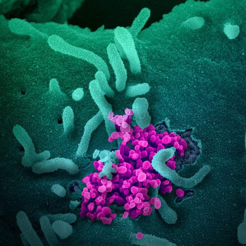
- Bad Bacteria: Cause diseases like strep throat and food poisoning.
Most bacteria help humans and are safe. A few can make people sick.
Bacterial Shapes and Groupings
Bacteria come in many shapes. They can be:
- Cocci: Round-shaped bacteria.
- Bacilli: Rod-shaped bacteria.
- Spirilla: Spiral-shaped bacteria.
Bacteria group in clusters, chains, or stay alone.
Gram Stain Classification
Bacteria are grouped by cell wall color after a special stain:
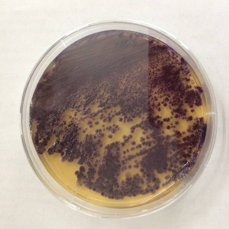
- Gram-Positive: Stain purple, have thick cell walls.
- Gram-Negative: Do not keep the purple stain, have thinner walls.
The Gram stain helps decide which antibiotics may work to treat infections.
Laboratory Protocols and Safety
When working with bacteria under a microscope, following proper laboratory protocols and ensuring safety are critical. These guidelines help maintain equipment, prevent contamination, and protect against potential hazards associated with handling bacterial cultures.
Cleaning and Maintenance of Equipment
Regular cleaning and maintenance of your microscope are essential. After each use:
- Turn off and unplug the microscope.
- Wipe lenses gently with lens tissue.
- Clean the stage and other surfaces with 70% ethanol.
- Cover the microscope with a dust cover when not in use.
This routine keeps your microscope in good condition, ensuring clear images and prolonged equipment life.
Safe Handling of Bacterial Cultures
Handling bacterial cultures requires caution. To ensure safety:
- Wear gloves and a lab coat.
- Sterilize tools like loops and swabs before and after use.
- Avoid eating, drinking, or touching your face in the lab.
- Dispose of used slides and coverslips in a biohazard container.
By following these practices, you reduce the risk of spreading bacteria and creating a hazardous environment.


