Introduction to Microscopy and Bacteriology
Microscopy is a fascinating science that brings the unseen world into view. It’s a method that uses microscopes to magnify tiny objects like bacteria, usually invisible to our naked eyes. Bacteriology is the study of bacteria, those miniature life forms surrounding us in countless numbers. Although you can’t spot germs with just your eyes, microscopes make it possible.Learn about Germs under a microscope.
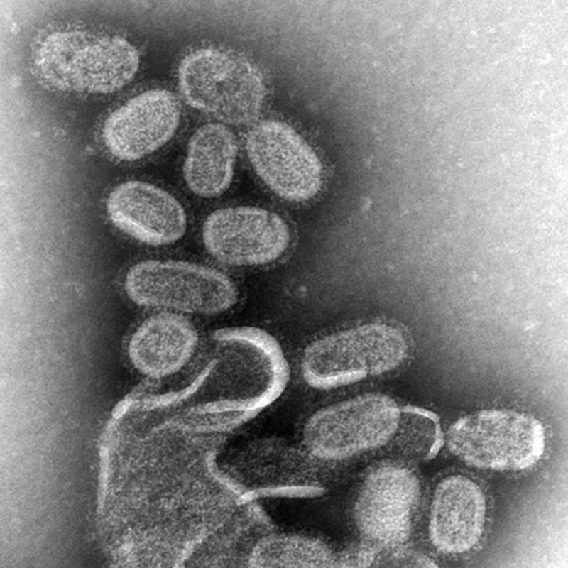
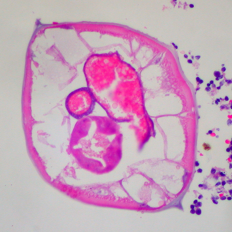
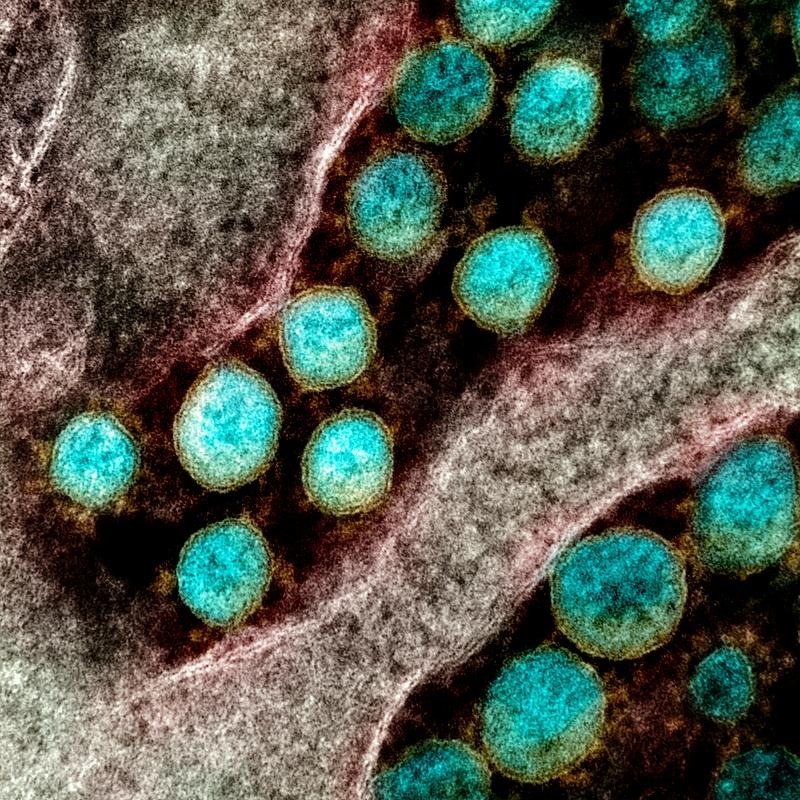
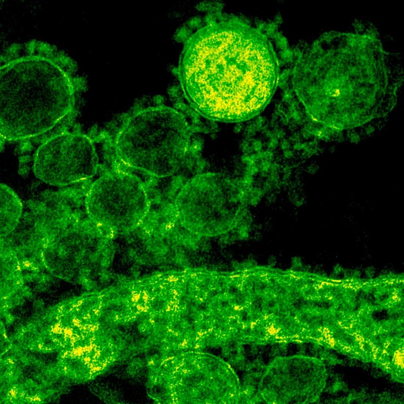
When you place germs under a microscope, their hidden features stand out. These microbes are around 0.2 to 8 micrometers in size, making it essential to use high-powered lenses to see them. Shapes vary, with spheres, rods, and spirals among the most common. With a microscope, minute bacterial details pop into view after some careful preparation and staining.
Discovery and research on bacteria are crucial. They affect our environment, health, and science. Some cause illness, while others play a part in curing diseases or breaking down waste. By studying germs under a microscope, scientists can understand and control their effects better.
To start exploring this micro world, you first need a quality microscope. Different types serve various purposes in revealing bacteria. A compound microscope is most common for its multiple magnifying lenses. A phase-contrast microscope provides detail without the need for stains. Both are key tools in bacteriology.
Whether in a professional lab or a school classroom, using a microscope opens a door to the tiny wonders all around us. Viewing germs under a microscope not only aids in research but ignites curiosity and a deeper respect for the complexity of life at a microscopic level.
Preparing Your Microscope for Viewing Germs
Viewing germs under a microscope requires a meticulous setup. Begin by choosing a clean, stable surface for your microscope. Ensure it has a power source, whether from an outlet or charged batteries. Next, adjust the lighting to a level that is easy on your eyes.
Cleanliness is essential. Wipe down your microscope’s lenses with proper lens paper to prevent scratches and remove any dirt. Set the lens to the lowest magnification first. This helps locate your sample on the slide.
Your microscope’s focus knobs are crucial. Start with the coarse adjustment to bring your specimen into view. Then, use the fine adjustment for a sharp, crisp image.
When everything is set, you’re ready to mount your slide. Secure it firmly on the stage to avoid slips. By following these steps, you’re well-prepared for observing germs under a microscope. The goal is to achieve a clear view of these tiny life forms. Remember, take your time and be patient to ensure accurate observations.
Collecting Samples to View Under a Microscope
Before diving into the world of microorganisms, you must collect a sample to put under the microscope. Proper sample collection is vital for observing germs under a microscope accurately. Here’s how you can gather samples effectively:
- Identify the Sample Source: Start by choosing where to collect from. Potential sources include water, soil, surfaces, or organic material.
- Sterilize Your Tools: Use a sterilized loop, swab, or pipette to prevent contamination. This ensures that only the germs from your sample are observed.
- Collect the Sample: Gently gather the material. If it’s a liquid, a slight suction with a pipette is enough. For solids, lightly rub the swab over the material.
- Transfer to a Slide: Carefully place your sample on a clean microscope slide. Ensure the slide is free from any dust and fingerprints.
- Prepare for Staining: If you’re using a stain to enhance visibility, make sure the sample is in the right condition for the stain to adhere properly.
While collecting samples, it’s crucial to be methodical. Any mistakes in collection could lead to misleading observations. Make notes of where and how the sample was collected. This information can be important later when identifying the germs under the microscope. Remember, always handle samples with care and use appropriate safety equipment to protect yourself from any potentially harmful germs.
Applying Stains for Enhanced Visibility of Germs
When observing germs under a microscope, using stains is key. Stains highlight the germs, making them easier to see. In this section, we’ll guide you through staining your samples for the best results.
- Choose the Right Stain: Methylene blue and crystal violet are common choices. They bind well with the bacterial components.
- Apply the Stain: Place a drop on your sample. Use a sterile loop to spread it gently.
- Let it Dry: Wait for the stain to dry. This step is crucial for clear results.
- Rinse if Needed: Some stains require rinsing. If so, gently wash the slide with water.
- Air Dry the Slide: Before observation, ensure the slide is completely dry.
Using a stain can turn transparent germs into visible, colorful specks. This makes detecting and identifying the shapes and arrangements of germs easier. Keep a steady hand and a patient mind when applying stains. This ensures a neat sample with well-defined germs under the microscope.
Step-by-Step Guide to Observing Germs Under High Magnification
To witness the intricate details of germs under a microscope, follow this step-by-step guide designed with simplicity and clarity in mind.
- Start with Low Magnification: Begin observing at lower power magnification. This helps find the area of interest without excessive detail.
- Use Coarse Adjustment: After locating your sample, the coarse focus knob brings it roughly into view. Turn it slowly to avoid damage to the slide.
- Switch to High Magnification: Move to a high-powered objective lens carefully. This is usually the 40x or 100x lens, ideal for detailed observation of germs.
- Fine-Tune Your View: With high power selected, use the fine adjustment knob. Make minor tweaks until the germs come into sharp focus.
- Slide Positioning: Sometimes, increasing magnification requires repositioning the slide. Do it gently to keep the germs in the field of view.
- Watch for Details: Now, look for the characteristic shapes and motions. Germs under a microscope can appear as dots, rods, or spirals, each hinting at a different type.
- Be Patient: High magnification often means a smaller field of view. Move the slide slowly to explore different areas and find your germs.
Remember, capturing the elusive beauty of germs under a microscope requires practice. Approach each step with care, maintaining focus on hygiene and safety. With patience and precision, the microscopic world will unfold, revealing the spectacular details of these tiny life forms.
Adjusting Magnification Levels for Clear Bacterial Observation
To see the tiny details of germs under a microscope, adjust the magnification carefully. Begin with the lowest setting, usually a 4x or 10x lens. This enlarges the sample enough to find the area you want to study. Once located, you can switch to high magnification levels. Use a higher-powered lens like 40x or 100x to zoom in on the bacteria.
High magnification brings germs into clear view. The higher the magnification, the smaller the field of view becomes. So you may need to move the slide a little. This helps keep the germs within your sight. When you change lenses, refocus using the fine adjustment knob. Small, precise turns of this knob will sharpen the image. At 1000x magnification, some microscopes allow you to see even finer details.
As you switch to higher magnification, increase the light if required. More light can improve the clarity of germs under a microscope. Adjust the diaphragm to regulate light. This controls the amount and focus of light that reaches the specimen.
Observing bacteria with the right magnification reveals their true shapes. Whether they are spheres, rods, or spirals, clear observation depends on this adjustment. With patience and the right settings, you’ll see germs formerly invisible to the naked eye.
Types of Microscopes Suitable for Viewing Germs
Choosing the right type of microscope is crucial for viewing germs effectively. Let’s look at what options are available:
Compound Microscopes
This common type has multiple lenses, providing high detail and clarity. Compound microscopes amplify germs using an eyepiece and objective lenses. The high magnification levels, typically between 40x and 100x, allow users to see bacteria clearly.
Phase-Contrast Microscopes
These microscopes enhance the visibility of transparent germs. They don’t require stains, so germs retain their natural state during observation. Phase-contrast microscopes use special optics to distinguish the different refractive indexes in germs.
In a nutshell, to view germs under a microscope, you need a tool that can magnify the small and the invisible. The compound microscope is the go-to for its multiple magnification capabilities. For live, unstained germs, the phase-contrast microscope might be your choice, allowing you to see germs in action without altering them. Make sure to choose what suits your needs for clarity and detail.
Documenting Your Observations of Germs
Once you have detected germs under a microscope, recording your findings is vital. Here are some tips to document your observations accurately:
- Take Notes: Write down important details. Note the magnification level, sample source, shapes, and arrangements.
- Draw What You See: A quick sketch can help later when comparing with textbooks or databases.
- Take Photographs: If your microscope has a camera, use it. Photos are precise records of your findings.
- Label Carefully: On your slides or in your notes, label everything. Include the date, sample type, and stains used.
- Create a Logbook: Keep a dedicated record book. Enter every session’s data for future reference.
As you document, be precise and thorough. Your records will be useful for future studies or sharing with others. Remember, attention to detail now can lead to significant discoveries later.
Safety and Hygiene Practices in Microscopic Observations
Working with microscopes and observing germs poses potential risks. It’s essential to follow safety and hygiene protocols to prevent contamination and ensure safe handling. Here’s a list of best practices:
- Wear Protective Gear: Always wear gloves and a lab coat. Eye protection is also crucial.
- Disinfect Surfaces: Before and after use, wipe down all surfaces with a disinfectant.
- Handle Slides with Care: Use forceps or gloves to avoid direct contact with microscope slides.
- Proper Sample Handling: Collect and dispose of samples in a manner that prevents the spread of germs.
- Avoid Eating or Drinking: Never consume food or beverages in a lab environment.
- Wash Hands Regularly: Before and after handling samples, thoroughly wash your hands.
- Use Caution with Stains: Handle all stains with care, as some may be harmful.
- Disposal of Waste: Dispose of all lab waste appropriately according to safety guidelines.
- Clean Equipment Properly: Follow the manufacturer’s instructions to clean and maintain your microscope.
By ensuring these practices, you minimize the risks and maintain a clean environment. Safety is vital, especially when dealing with germs that could be potentially harmful. Be mindful and diligent in your approach to microscopic observations.
The Role of Microscopy in Germ Research and Discovery
Microscopy is a critical tool in the world of germ research and discovery. By allowing us to view germs under a microscope, this technology enables scientists to dive deep into the microscopic universe. Below are some of the ways in which microscopy plays a pivotal role:
- Understanding Germ Behavior: By observing how germs move and interact under a microscope, researchers learn about their behavior. This can lead to important insights into how diseases spread.
- Identifying New Species: The high magnification levels of microscopes help discover new germ species previously unknown to science. Each discovery could hold the potential for new treatments or understanding of ecosystems.
- Antibiotic Resistance Study: Microscopy allows scientists to examine how certain germs become resistant to antibiotics. Observing these changes can help in developing strategies to combat resistance.
- Environmental Impact Analysis: Researchers study how germs under a microscope affect various environments. Microscopes help in monitoring germ populations and their effects on ecosystems.
- Medical Diagnoses Support: Diagnosing infections often depends on identifying the responsible germs. Microscopes make this process more accurate, supporting effective treatment.
- Vaccine Development: Understanding germ structure and function is essential in creating vaccines. Microscopes offer a close-up view, aiding vaccine research.
- Educating the Next Generation: Microscopes in classrooms expose students to the wonders of microbiology. They spark curiosity and interest in future scientists.
Microscopy transforms invisible germs into visible specimens that can be studied. It arms researchers with the ability to observe, identify, and understand germs on a level that impacts global health and science. In a way, microscopes serve as windows into a world that, although tiny, is immense in its significance. The knowledge gained through these observations continues to drive innovation and discovery across multiple scientific fields.


