Introduction to Blood Microscopy
Blood microscopy is a vital scientific technique. It reveals what the naked eye can’t see in blood. The method involves examining blood under a microscope to inspect its components closely.
When blood is placed under microscopic lenses, its elements become visible. You can see red and white blood cells, platelets, and plasma. Each has a role in bodily functions and health. Now, with advanced microscopy, we can study these cellular structures in detail.
The procedure starts with a well-prepared blood slide. It’s stained to highlight different parts. Microscopists then scrutinize it to assess blood health and diagnose conditions. This process unveils signs of disorders that might otherwise go unnoticed.
Blood under microscope exams help clinicians diagnose countless conditions. They range from nutritional deficiencies to immune system disorders. The technique is also essential for research, unlocking secrets of blood-related diseases.
Throughout this blog, we will dive deeper into each blood component and how they appear under a microscope. We’ll also explore blood preparation techniques and discuss the impact of technological advancements on blood microscopy.
The Components of Blood Revealed Under Microscope
When we look at blood under microscope, we unveil a complex world. Each component plays a crucial role in our health. Here’s what you can expect to see:
Red Blood Cells
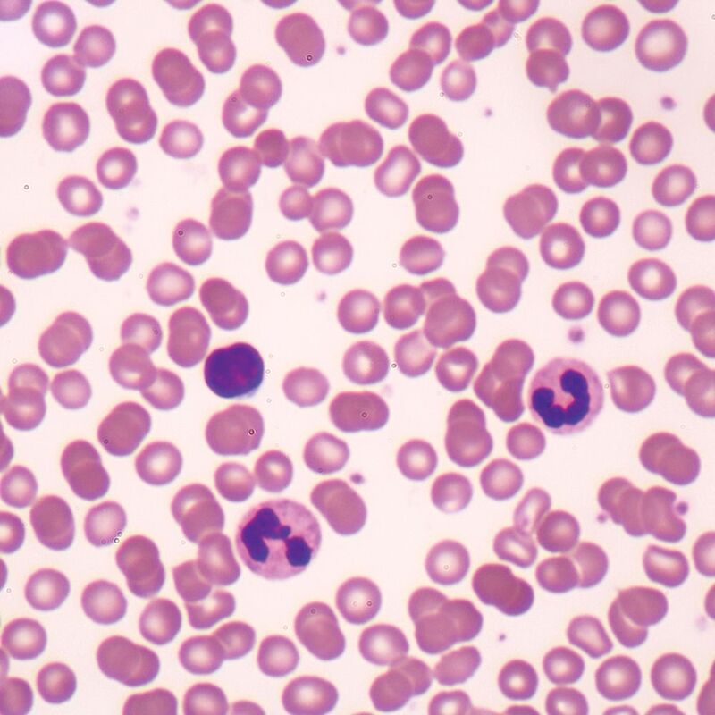
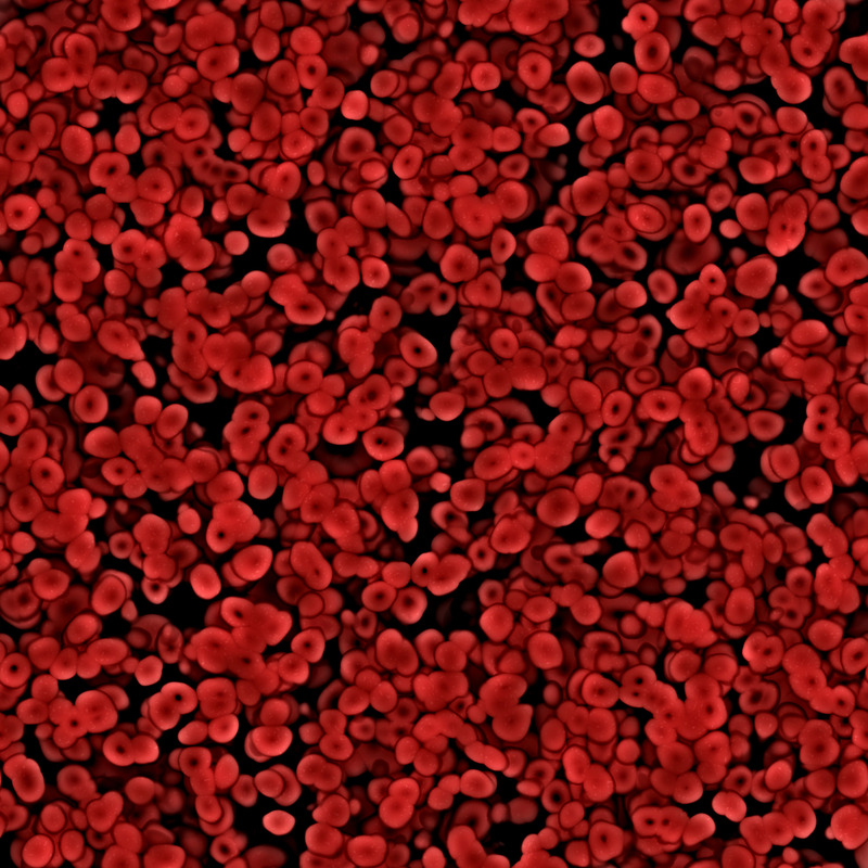
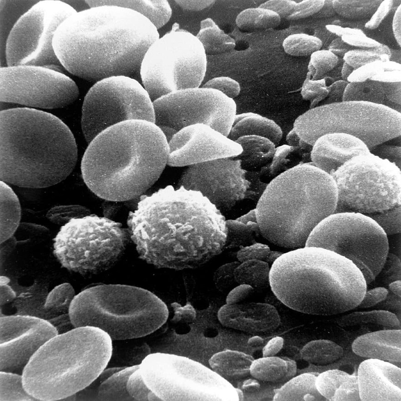
These are the most abundant cells in our blood. They look like tiny, flexible discs with a dip in the center. Red blood cells carry oxygen from our lungs to the rest of our body. When you view them under a microscope, their reddish hue is unmistakable.
White Blood Cells
These cells are the warriors of our immune system. They come in various shapes and sizes, often appearing larger than red blood cells. White blood cells defend our body against infections. Under a microscope, you can sometimes see them actively hunting down bacteria.
Platelets
Platelets are small fragments that play a key role in blood clotting. They work to prevent excessive bleeding when we get hurt. Under high magnification, you’ll notice they are smaller than red and white cells and have no standard shape.
Plasma
Plasma is the liquid part of blood, a yellowish fluid. It carries blood cells and nutrients throughout our body. In a microscope slide, plasma creates the background in which other blood components float.
Each element of blood has distinctive features and essential functions. Viewing them under a microscope not only helps understand our body better but also aids in diagnosing diseases. In the following sections, we will explore how these components change in common blood disorders.
The Process of Preparing a Blood Slide
Preparing a blood slide is a meticulous task that requires precision and patience. It’s a critical step before examining blood under microscope. Here we’ll go over the basic steps involved in creating a usable blood slide for microscopy.
- Collection: Blood is usually collected through a finger prick or venous draw, using sterile techniques to avoid contamination.
- Smearing: A drop of blood is placed on a clean microscope slide. Using another slide, it is spread thinly across the surface to create a ‘blood smear’.
- Drying: The smear is left to air dry. Hastening this process with heat can warp cells, so patience is key.
- Fixing: Once dry, the smear is often fixed with methanol. This preserves the blood cells’ structure.
- Staining: Specific stains, like Wright’s or Giemsa stain, are applied. These bring out details in the cells that wouldn’t be visible otherwise.
- Rinsing: Excess stain is gently washed off, not disturbing the cells.
- Drying again: The slide is air-dried once more. It must be completely dry before viewing.
- Viewing under a microscope: Finally, the slide is placed under the microscope. Different magnifications are used to observe various components.
Each step is vital to ensure a clear view of the blood under microscope. A well-prepared slide reveals intricate details of blood cells, necessary for accurate analysis. In the next section, we’ll examine how certain common blood disorders can alter the appearance of blood components under microscopic examination.
Common Blood Disorders and Their Appearance Under the Microscope
Blood disorders often impact the appearance of blood under microscope. Certain conditions can modify the shape, size, or number of blood cells. We will look at a few common blood disorders and how they change what we see on a slide.
Anemia
In cases of anemia, red blood cells look paler due to low hemoglobin. They may also appear misshapen or varied in size. Anemic blood under microscope shows a clear reduction in the overall red cell count.
Leukemia
Leukemia shows a high number of white blood cells, often immature. These cells may look irregular and out of proportion compared to healthy cells. When viewing blood under microscope, the crowded white cells become evident.
Thrombocytopenia
In thrombocytopenia, there’s a noticeable decrease in platelet count. This can be seen under a microscope as a scarcity of the small cell fragments. Sometimes, the existing platelets may seem larger than normal.
Hemophilia
While hemophilia does not change blood cell appearance, it affects clotting factor levels. This condition might not be directly observable under microscope without specific tests. But, experts look for signs like unusually large blood clots or clumps of cells.
Each disorder leaves its unique imprint on blood, which specialists can decipher. Understanding how these conditions alter the look of blood under microscope aids in prompt and precise diagnosis.
Technological Advances in Blood Microscopy
The field of blood microscopy has seen remarkable advancements in technology. These innovations have vastly improved the clarity and detail of what we can observe when looking at blood under microscope.
Enhanced Imaging Techniques
New imaging techniques offer sharper and more detailed views of blood cells. High-resolution microscopes now have the ability to magnify cells up to a thousand times or more. This lets us see finer details within the cells, such as the structure of the cell membrane.
Digital Microscopy
Digital microscopy is a game-changer. It allows us to capture and store images of blood samples on computers. With this technology, we can easily share these images for teleconsultation or remote diagnosis. Additionally, software can analyze the images and highlight areas of interest.
Automated Cell Counters
Automated cell counters use flow cytometry to quickly count and categorize cells in a blood sample. These devices have streamlined the process of identifying and quantifying blood cells, making it faster and more accurate.
Advanced Staining Methods
Staining techniques have improved, with more precise stains that bind to specific cell components. This specificity helps in distinguishing between different types of blood cells and spotting anomalies that could indicate disorders.
Fluorescence Microscopy
Fluorescence microscopy uses specific dyes that emit light when exposed to certain wavelengths. This makes particular elements within the blood ‘glow’, thus, easing the identification of structures like proteins or DNA within cells.
As we harness these technological advances, the potential for understanding and diagnosing blood-borne diseases continues to grow. Aided by these tools, researchers and clinicians can delve deeper into the microscopic world of blood and unearth findings that were previously unattainable.
Interpreting Blood Test Results from Microscopic Analysis
Interpreting blood test results involves understanding the microscopic details. Experts analyze blood under microscope to assess health and diagnose conditions. They compare observed cell shapes, sizes, and counts with normal ranges to identify abnormalities. Here’s a breakdown of the interpretive process:
- Evaluation of Red Blood Cells: A normal view shows uniform cells with central pallor. Variations might suggest anemia or other disorders.
- White Blood Cell Assessment: The number and type of white cells are scrutinized. Abnormalities can indicate infections or blood cancers like leukemia.
- Platelet Count: An accurate platelet count is critical. It helps evaluate clotting capabilities and diagnosing diseases like thrombocytopenia.
- Plasma Analysis: The plasma’s color and consistency give clues about hydration, liver function, and other metabolic aspects.
The accurate interpretation of blood slides is essential for patient prognosis. It guides treatment decisions and monitors the efficacy of interventions. As such, mastering the skill of reading blood under microscope is key for any hematologist or laboratory technician.
The Future of Blood Analysis and Microscopic Technology
As we envision the future of blood analysis, microscopic technology is advancing at an impressive pace. The evolution of this field promises more accurate diagnoses, real-time monitoring, and personalized treatment plans. Innovations are reshaping the landscape of hematology and patient care in several ways:
- Automation and AI Integration: Future microscopes may employ artificial intelligence (AI) to detect and diagnose blood disorders automatically. This can reduce human error and speed up lab workflow.
- Miniaturized Devices: Portable, handheld microscopes are becoming a reality. They could enable bedside blood analysis, making diagnostic testing accessible in remote areas.
- Nanotechnology: Researchers are experimenting with nanotechnology in blood microscopy. This could lead to detecting diseases at the molecular or even atomic level.
- Virtual Reality (VR) and Augmented Reality (AR): VR and AR could revolutionize training for professionals by simulating blood analysis scenarios.
- Personalized Medicine: As technology advances, we expect to see more tailored therapies based on individual blood cell analysis.
These advancements in microscopic technology herald a new era for blood examination. They show immense potential to enhance our understanding of blood-related health issues and the underlying mechanisms of diseases. As a result, we may witness significant improvements in patient outcomes and the efficacy of treatments. Watching blood under microscope will become not just a diagnostic procedure, but also a gateway to innovative healthcare solutions.


