Introduction to Compound Microscopes
A compound microscope is an essential tool in many scientific and medical settings. This type of microscope uses multiple lenses to achieve higher magnifications, allowing for detailed observation of microscopic structures. Compound microscopes amplify tiny details of specimens such as cells, microorganisms, and thin tissue slices.
The structure of a compound microscope includes several key components that work together to produce clear, magnified images. These components encompass both optical parts, responsible for magnification and visualization, and structural parts that provide stability and support.
In studying compound microscopes, it’s vital to understand how these parts function individually and in conjunction with each other. For instance, light from the microscope’s illuminator passes through the specimen. The objective lenses then magnify the image, which is further enlarged by the eyepiece or ocular lens for the observer to see.
Each part of the microscope has a specific role that contributes to the overall effectiveness of the microscope. Understanding these roles can enhance the user’s ability to effectively use the microscope for research, diagnostics, and educational purposes.
By grasping the basics of how a compound microscope is structured and functions, users can better troubleshoot issues, optimize the microscope’s performance, and achieve more accurate observations and results in their microscopic studies.

Key Structural Components of a Compound Microscope
Head (Body Tube) of the Microscope
The head, or body tube, is a critical part of the compound microscope. It is a long cylindrical structure that connects the eyepiece to the microscope’s objective lenses. It ensures the light path is aligned correctly, leading to a clear image in the eyepiece.
Microscope Base
The base acts as the microscope’s foundation. It provides stability and typically houses the illuminator or light source. Without a stable base, the microscope could not accurately focus on specimens.
The Arm and Its Significance
The arm serves as the microscope’s backbone. It supports the head, allowing for precise alignment of optical components. It is also the main handle when transporting the microscope, ensuring safe movements.
Core Optical Parts of a Compound Microscope
The core optical components are key for viewing details in specimens. These include the eyepiece or ocular lens, objective lenses, and the nosepiece. Each part plays a vital role in the magnification process, transforming the compound microscope into a powerful investigative tool.
Eyepiece (Ocular Lens) Functionality
The eyepiece is where you look in to see the magnified image. It usually magnifies the object 10 times (10x). This lens works with objective lenses to enhance the overall magnification of the microscope.
Objective Lenses and Magnification
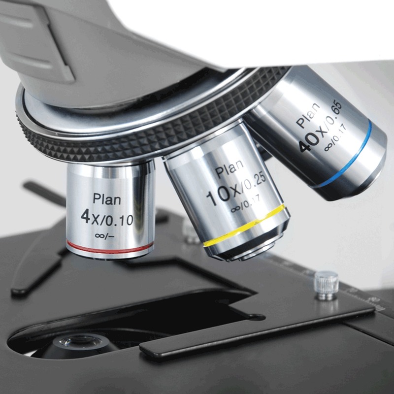
Objective lenses are crucial for primary magnification. They come in different strengths, providing varied levels of zoom. Typically, you’ll find 4x, 10x, 40x, and 100x magnification lenses on a microscope to view samples with greater detail.
Nosepiece and Its Role in Magnification
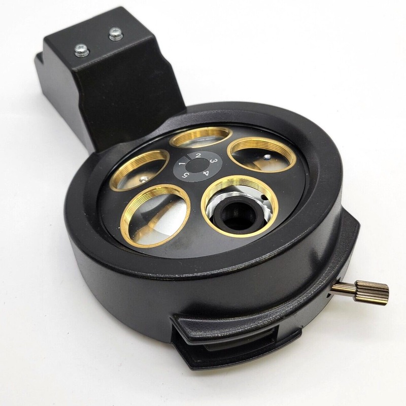
The nosepiece holds the objective lenses. It rotates to switch between lenses, changing the magnification power. This part ensures that the microscope can focus on specimens at different degrees of enlargement, allowing a comprehensive examination of microscopic details.

Focusing Mechanisms on a Microscope
Focusing the microscope is critical to getting sharp images of your specimen. This is where the adjustment knobs and stage controls come into play.
Fine and Coarse Adjustment Knobs
Two types of adjustment knobs help in focusing: the fine and the coarse knobs. The coarse adjustment knob lets you quickly bring the specimen into general focus. It moves the stage up and down rapidly. After, you use the fine adjustment knob for precise focusing, especially at high magnifications. It moves the stage slowly to sharpen the image.
Stage Controls and Stage Clips
Stage controls are knobs that shift the slide left or right, and forward or backward. This helps in scanning different areas of your specimen. Stage clips hold the slides in place, ensuring they stay still during observation. Together, these components work to keep the image focused and centered for detailed examination.
Illumination and Image Clarity
Proper lighting is crucial for clear images in microscopy. Let’s explore the parts that help in this regard.
The Integral Role of Illuminators
An illuminator is a microscope’s light source. It brightens the specimen, making details visible. Often found in the base, the illuminator can be a lamp or LED.
Condenser, Diaphragm, and Aperture
The condenser focuses light onto the specimen. The diaphragm, sitting above it, adjusts light intensity. Aperture, the stage hole, lets light reach the specimen.
Importance of Light and Brightness Adjustment
Adjusting light is key for image quality. Controls for light and brightness let you fine-tune the illumination. This ensures you get the best possible image clarity.
Difference Between Stereo and Compound Microscopes
Stereo and compound microscopes serve distinct purposes and are suited for different types of observations.
Magnification and Resolution in Microscopes
Stereo microscopes provide lower magnification but a three-dimensional view, ideal for observing larger, thicker specimens. Compound microscopes offer higher magnification and resolution, crucial for examining microscopic entities.
Applications of Stereo vs. Compound Microscopes
Stereo microscopes are used in fields such as entomology and circuit board analysis. Compound microscopes are essential in cellular biology and medical diagnostics.

Advanced Microscope Technologies
The landscape of microscopic examination has broadened with the advent of advanced microscope technologies. These innovations provide superior magnification, resolution, and insight into the cellular and molecular world. Here, we delve into notable advancements such as electron microscopy, fluorescence microscopy, and phase contrast microscopy, highlighting their functionalities and applications.
Electron Microscopy Innovations
Electron microscopy stands out due to its incredible magnification powers, allowing scientists to view structures at the nanometer scale. This technology uses beams of electrons instead of light which results in much higher resolution images. There are two main types: Transmission Electron Microscopy (TEM) and Scanning Electron Microscopy (SEM). TEM helps in viewing the internal composition of cells and molecules, while SEM provides a 3D topographical surface analysis of specimens. These capabilities make electron microscopy indispensable in material science, biology, and forensic applications.
Fluorescence and Phase Contrast Microscopy
Fluorescence microscopy involves using high-intensity light to illuminate dyes that are attached to specific cell and tissue components. The dyes emit light upon excitation, which is then magnified. This technique is pivotal in studying cellular structures, functions, and locating proteins.
Phase contrast microscopy, on the other hand, enhances the contrast in transparent and colorless specimens without requiring staining. It converts phase shifts in light passing through a specimen into brightness changes in the image. It’s particularly useful in biomedical research for observing live cells and organisms, providing clear images of intracellular processes.
These advanced technologies aid significantly in scientific breakthroughs, offering detailed views into the micro-world not visible to the naked eye. Understanding and utilizing these technologies enhances our ability to analyze biological specimens, perform complex biomedical research, and develop medical diagnostics.
Conclusion and Importance of Understanding Microscope Parts
In conclusion, recognising each part of a microscope has great significance. By understanding the various components and their functions, users can utilize the instrument more effectively. This knowledge is beneficial for troubleshooting, optimizing use, and achieving precise results.
The head of the microscope links the ocular lens and the objective lenses. This ensures clear viewing. The sturdy base and arm support the microscope, allowing for stable and safe handling. Properly using the eyepiece, objective lenses, and the rotating nosepiece leads to varied magnification levels. Thus, one can examine items in detail.
Knowing how to adjust the focus with the coarse and fine knobs is key. It ensures sharp images. The stage controls and clips aid in examining different areas of a slide. The illuminator, condenser, diaphragm, and aperture all work to make samples bright and visible. Proper light adjustment means clear images for observation.
Comparing stereo and compound microscopes highlights their unique purposes. Stereo microscopes are for larger, 3D viewing, while compound microscopes offer higher magnification for small, detailed study. Both types suit different scientific needs.
Lastly, advanced technologies like electron, fluorescence, and phase contrast microscopes extend our research capabilities. They equip us with new ways to explore cells and materials at the microscopic level.
Overall, comprehensive knowledge of microscope parts enhances scientific investigation, supporting new discoveries and advancements in various fields.


