Introduction to the Compound Light Microscope
The compound light microscope, an essential tool in biological research and education, amplifies small objects using lights and lenses. Unlike magnifiers with a single lens, the compound variant achieves higher magnification through a series of optical components. These include the objective lenses near the specimen and the eyepiece, through which the viewer observes.
Its ability to provide detailed views of tiny entities, like cells and bacteria, makes it crucial in laboratories. Not just limited to researchers, school students often encounter the compound light microscope in science classes, learning about life on a micro-scale.
The compound light microscope operates straightforwardly. Light from a built-in source travels through a transparent specimen. As it ascends, it passes through the objective lenses, forming a magnified image that the eyepieces further enhance. This generates a clear, enlarged picture of the examined sample.
In the forthcoming sections, we’ll delve deeper into the history, structure, and working principle of the compound light microscope. We’ll also explore its many applications, benefits and limitations, and how it compares to other types of microscopes. Understanding these aspects will help us appreciate the compound light microscope’s value in scientific discovery and education.
Historical Development of the Compound Microscope
Tracing the origins of the compound light microscope leads us back to the late 16th century. Dutch spectacle-maker Zacharias Janssen is frequently credited with creating the first compound microscope around 1590, though this claim lacks strong documentary evidence. Regardless of its disputed invention, it’s widely accepted that these early devices consisted of a tube with lenses at both ends, significantly improving magnification compared to the single-lens simple microscope of the time.
The development of the compound microscope evolved through the 17th century with contributions from notable figures, such as Galileo, who enhanced the design, and Antonie van Leeuwenhoek, who made improvements that allowed for the detailed observation of bacteria and cells, a monumental step in microbiology. The subsequent centuries saw the compound microscope become increasingly refined. Innovations included better lens-grinding techniques and the introduction of achromatic lenses to reduce chromatic aberration—a distortion of color at the fringes of an image.
By the 19th century, the compound microscope’s applications had expanded far beyond its initial use. Carl Zeiss, a prominent optician, played a crucial role in the microscope’s development. His collaboration with physicists and chemists led to improved optics and the expansion of the compound microscope’s capabilities.
Throughout the 20th century and into the contemporary era, advancements in technology have turned the compound microscope into a sophisticated instrument. Electron microscopy and digital imaging have broadened the boundaries of what can be observed at the cellular level. Despite these technological leaps, the compound light microscope remains a fundamental tool, whose historical development underpins its modern-day value in education, research, and a plethora of scientific fields.
Key Components and Functionality
A compound light microscope consists of various elements that are crucial for its operation. Let’s explore these components, segmenting them into optical and non-optical parts.
Optical Parts of the Compound Microscope
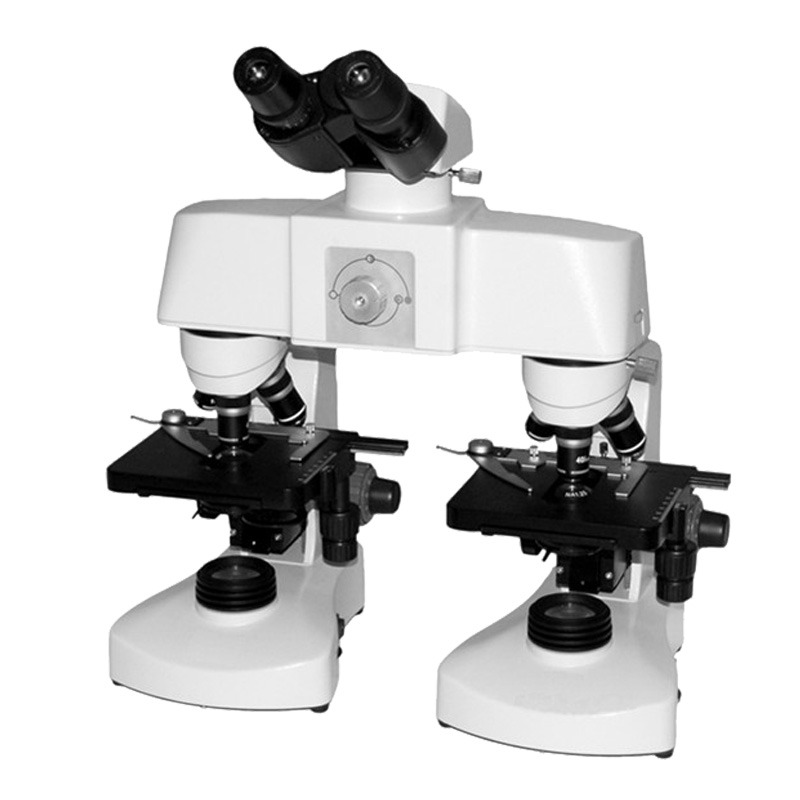
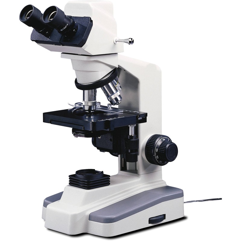
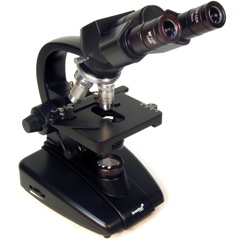
The optical components of the compound light microscope are vital for magnification and image formation. The objective lenses are positioned close to the specimen to first magnify the image. Next, the ocular lens, or eyepiece, enlarges this image further to enable detailed observation. The compound microscope typically includes three objective lenses—low power, high power, and oil immersion. Each lens is used based on the needed magnification.
Another essential part is the diaphragm, which controls the amount of light that reaches the specimen. Adjusting the diaphragm optimizes the light and improves the image quality. The condenser focuses the light onto the sample, which is crucial for a clearer view. A reflector or mirror directs light from the microscope’s light source through the diaphragm.
Non-optical Parts of the Compound Microscope
Non-optical components provide structural support and precision handling. The base supports the whole microscope, often accompanied by a sturdy arm used to carry the microscope safely. Connected to the base is the pillar, which holds the arm and allows the microscope’s head to tilt for comfortable viewing through the inclination joint.
The specimen is placed on the stage, typically equipped with clips to hold slides in place. The body tube connects the ocular and objective lenses. Focusing is achieved through the coarse and fine adjustment knobs. These knobs adjust the distance between the objective lens and the specimen to bring the image into sharp focus.
Each part of the compound light microscope plays a specific role, collectively contributing to its functionality and efficiency in various scientific settings.
The Principle of Operation: How a Compound Microscope Works
The compound light microscope employs a straightforward yet precise process to magnify objects. It begins with light originating from the microscope’s own light source. This light passes through the transparent sample placed on the stage. As the light travels upward, it encounters the objective lenses. These lenses magnify the image in steps, starting with the low power lens and progressing to higher powers if needed. The high power and oil immersion lenses offer greater magnification for detailed viewing.
A real image is formed as the light passes through the objective lens and this image is further enlarged by the ocular lens, or eyepiece, as we observe it. What we see through the eyepiece is a virtual image, an enlarged and inverted version of the real one. This system provides a bright, clear view due to the alignment of the lenses along with effective light management.
To achieve this, the microscope uses the diaphragm to control the light’s intensity and the condenser to focus it on the specimen. Adjustments can be made easily to ensure the lit area is optimal for viewing the sample. The precise positioning of the lenses and the ability to control light allows scientists and students to see structures which would otherwise be invisible to the naked eye. Collectively, these functions enable the compound light microscope to reveal a micro-world full of details and insights.
Advantages and Disadvantages
When considering a compound light microscope, understanding its strengths and limitations is critical.
Advantages
- High Magnification Levels: The compound light microscope offers significant magnification. It uses multiple lenses to magnify objects, making detailed study possible.
- Built-In Light Source: These microscopes come with their own light sources. This feature improves visibility and clarity of specimens.
- User-Friendly: They are generally easy to handle and operate. This makes them suitable for both students and professionals.
- Detailed Observations: Capable of revealing minute details in specimens. This is essential in fields such as microbiology and pathology.
Disadvantages
- Limited Magnification: Despite high levels, there’s a cap to how much you can magnify.
- Size Restrictions: Samples must be thin and translucent for the light to pass through.
- Complexity in Maintenance: Requires careful handling and regular maintenance to keep functioning properly.
- Cost: High-quality compound microscopes can be expensive due to their intricate design and functionality.
In summary, the compound light microscope is a powerful tool for scientific exploration, offering detailed views of tiny entities. However, users must weigh its high-cost and maintenance demands against its benefits.
Practical Applications of Compound Microscopes
Compound light microscopes have various uses in diverse fields. Their ability to magnify small objects makes them indispensable in science and industry. In this section, we’ll outline some of the key applications of compound light microscopes.
- Education: Compound microscopes introduce students to the wonders of the microscopic world. From high schools to universities, they are crucial tools in biology and science classes.
- Medical Research: Researchers rely on these microscopes to study cells, tissues, and microorganisms. They help in diagnosing diseases and understanding cellular structures.
- Pharmaceuticals: The development of new drugs often requires detailed analysis of chemical compounds. This is where compound microscopes play a vital role.
- Material Science: Scientists use them to examine material surfaces at the micro level. This helps in studying the properties of metals, polymers, and ceramics.
- Forensics: In forensic science, compound microscopes analyze samples like fibers, hair, or body fluids. They aid in solving crimes by providing vital clues.
- Environmental Science: Microscopes analyze soil and water samples. They can detect pollutants or microorganisms affecting the ecosystem.
- Food Industry: Quality control is vital, and microscopes check for contaminants in food. They ensure safety and compliance with health regulations.
- Electronics: They assist in inspecting circuit boards and components. Manufacturers use them to ensure quality at the micro-level in electronics.
Each application capitalizes on the compound light microscope’s ability to reveal details unseen by the naked eye. They are key tools in advancing our knowledge and improving many aspects of daily life.
Comparison With Other Microscope Types
When investigating microscopy tools, understanding their differences is essential. Here, we break down how a compound light microscope stands against other types.
Simple vs. Compound Microscopes
Simple microscopes use a single lens to magnify objects. They are suitable for basic viewing. Compound microscopes, by contrast, utilize multiple lenses. This achieves higher magnification and detail, ideal for scientific and educational purposes.
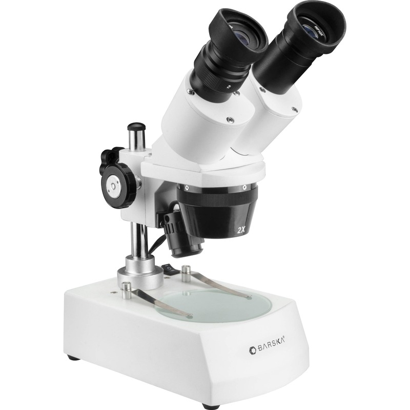
Stereomicroscopes vs. Compound Microscopes
Stereomicroscopes offer three-dimensional imaging. They use two optical paths to create depth perception. Compound microscopes, however, provide two-dimensional views. Their structure allows intense magnification but lacks depth. This makes them better for detailed cellular studies.
Each type of microscope has its place depending on the level of detail and dimension required. The choice between them depends on the specific requirements of the task at hand.
Maintenance and Care for Compound Microscopes
Proper maintenance and care of compound light microscopes are essential for their longevity and reliability. Here are some simple yet important guidelines to follow:
- Handle with Care: Always carry a microscope using its arm and base to avoid damage.
- Clean Regularly: Wipe lenses with lens paper and use a soft brush for dust.
- Cover When Not in Use: Protect your microscope from dust by covering it after each use.
- Avoid Touching Lenses: Fingerprints can blur the view, so handle lenses by their edges.
- Store Properly: Place the microscope in a dry, cool place away from direct sunlight.
- Check the Light Source: Ensure the built-in lighting works well and replace bulbs as needed.
- Inspect for Damage: Look for any signs of wear or damage, and get repairs done promptly.
- Calibrate: Regularly check that your microscope is properly aligned and calibrated.
Following these steps will help ensure that a compound light microscope continues to function effectively, allowing for clear and accurate observations in various applications.


